. 2014 Nov;10(11):884-91.
doi: 10.1038/nchembio.1670.

Energy landscapes of functional proteins are inherently risky

Affiliations

Energy landscapes of functional proteins are inherently risky

Anne Gershenson et al. Nat Chem Biol. 2014 Nov.

Abstract

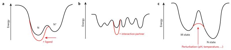
Evolutionary pressure for protein function leads to unavoidable sampling of conformational states that are at risk of misfolding and aggregation. The resulting tension between functional requirements and the risk of misfolding and/or aggregation in the evolution of proteins is becoming more and more apparent. One outcome of this tension is sensitivity to mutation, in which only subtle changes in sequence that may be functionally advantageous can tip the delicate balance toward protein aggregation. Similarly, increasing the concentration of aggregation-prone species by reducing the ability to control protein levels or compromising protein folding capacity engenders increased risk of aggregation and disease. In this Perspective, we describe examples that epitomize the tension between protein functional energy landscapes and aggregation risk. Each case illustrates how the energy landscapes for the at-risk proteins are sculpted to enable them to perform their functions and how the risks of aggregation are minimized under cellular conditions using a variety of compensatory mechanisms.


Figures

Figure 1
Figure 1. Schematic two-dimensional functional energy landscapes
(a) Many proteins sample a near-native state (N*) to open a binding site and interact productively with a ligand. Formation of the complex with ligand stabilizes the protein in its native state (N). (b) IDPs have sequences that disfavor a unique folded state and instead lead to many conformational possibilities of nearly equivalent energy. Interaction(s) with partners leads to stabilization of the state(s) that have the capacity to bind the partner with the highest affinity, thus shifting the energy landscape in the presence of partner(s) to one or only a few more stable states. (c) The magnitude of barriers on energy landscapes has a profound impact on protein function. A state that is not the thermodynamically most stable state (here M) may be long lived because it is separated from the most thermodynamically stable state (N) by high-energy barriers. The barriers may be reduced in amplitude by various triggers, whether environmental or protein-protein interactions.
Figure 2
Figure 2. CRABP1 exemplifies the structure and dynamics of the iLBP family
(a) Structure of holo-CRABP1 (ref. 104) (Protein Data Bank (PDB) code 1CBR) showing the helical portal domain, proposed to dynamically open to allow ligand entry and exit, (light orange ellipse), and several regions implicated in early folding events in CRABP1 (ref. 22) (green). (b) Regions of CRABP1 found to be sequestered in the cores of aggregates formed either in vitro or in E. coli cells are shown in orange. Note that the early folding events may provide a potential protective mechanism to offset the risk of aggregation during folding of apo-CRABP1. (c,d) Comparison showing increased dynamics of CRABP1 in the apo form (c) relative to holo-CRABP1 (d). The width of the polypeptide chain depicts the rate of exchange of backbone amides with solvent water, a clear indicator of chain dynamics. Panel c is modified with permission from a figure in reference 28. Copyright 2000, American Chemical Society.
Figure 3
Figure 3. Functional and nonfunctional serpin conformational gymnastics
(a) On the left is shown the active serpin conformation with the solvent-exposed RCL (red), sheet A (yellow) and the shutter, which must open to allow RCL insertion into sheet A. A target protease is shown as a blue space-filling structure as it interacts with the RCL in an initial encounter complex. On the right is shown the conformational transition required for mechanical inactivation of target proteases. (PDB codes 1OPH and 1EZX on the left and right, respectively). (b) Serpins can form multimers and polymers in vitro and in vivo. Possible interactions include, from left to right: addition of the RCL to the edge of a β sheet as observed in the antithrombin III dimer (PDB code 1E05 (ref. 107)), partial insertion of the RCL into sheet A of an adjacent monomer and domain swaps.
Figure 4
Figure 4. Folding landscape of β2m and its assembly with the MHC-1 heavy chain
(a) Schematic free energy landscape of β2m monomer folding and aggregation. Φ1 and Φ2 indicate the increase of native intramolecular interactions during folding and non-native intermolecular interactions during aggregation, respectively. The unfolded protein with cis-Pro32 and trans-Pro32 are denoted UC and UT; the intermediate ensembles are denoted as IC, which is populated in vanishingly small amounts, and IT, which is ~5% populated; and the native state is N. The fibril is indicated as Fib. (b) Structure of β2m showing cis-Pro32 (green), Trp60 (blue) and the disulfide bond (C23–C80) (sticks) (PDB code 2XKS)). The regions involved in binding to the MHC1 heavy chain are shown in orange on the surface. Panel a is reproduced from ref. 64.
Figure 5
Figure 5. Competition between ubiquitin binding and aggregation for the Josephin domain of Atx3
A surface representation of Josephin (PDB code 2JRI) is shown in white with exposed hydrophobic surfaces in green. When in the presence of the natural partner ubiquitin (Ub; the two binding ubiquitins are shown as blue traces), the Josephin binding sites are saturated by the interaction and protected from aggregation. When interaction is impaired, for instance by polyQ expansion, which alters the affinity for the interactor, the two sites promote aggregation, as shown in the right panel. Adapted from ref. 15 with permission from Elsevier.

References

    1. Jenik M, et al. Protein frustratometer: a tool to localize energetic frustration in protein molecules. Nucleic Acids Res. 2012;40:W348–W351. - PMC - PubMed
    1. Sutto L, Latzer J, Hegler JA, Ferreiro DU, Wolynes PG. Consequences of localized frustration for the folding mechanism of the IM7 protein. Proc. Natl. Acad. Sci. USA. 2007;104:19825–19830. - PMC - PubMed
    1. Zheng W, Schafer NP, Wolynes PG. Frustration in the energy landscapes of multidomain protein misfolding. Proc. Natl. Acad. Sci. USA. 2013;110:1680–1685. - PMC - PubMed
    1. Knowles TP, Vendruscolo M, Dobson CM. The amyloid state and its association with protein misfolding diseases. Nat. Rev. Mol. Cell Biol. 2014;15:384–396. - PubMed
    1. Uversky VN. Intrinsic disorder in proteins associated with neurodegenerative diseases. Front. Biosci. (Landmark Ed.) 2009;14:5188–5238. - PubMed

Publication types

LinkOut - more resources