. 2014 Dec 5;13(12):5347-61.
doi: 10.1021/pr500588c. Epub 2014 Oct 31.

Proteomic and biochemical analyses show a functional network of proteins involved in antioxidant defense of the Arabidopsis anp2anp3 double mutant

Affiliations

Proteomic and biochemical analyses show a functional network of proteins involved in antioxidant defense of the Arabidopsis anp2anp3 double mutant

Tomáš Takáč et al. J Proteome Res. .

Abstract

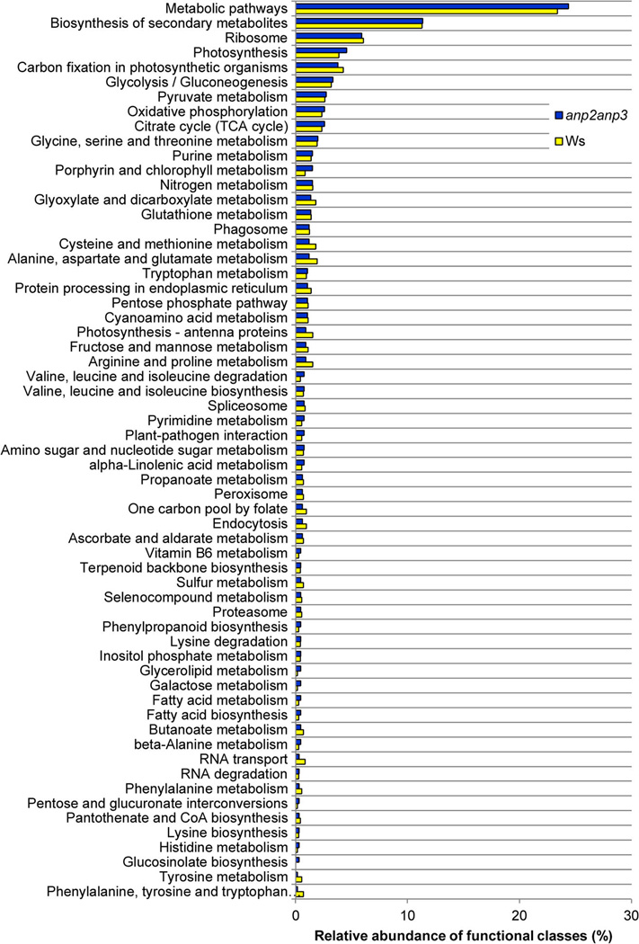
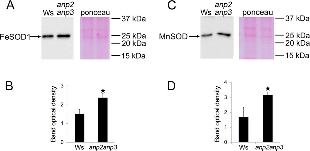
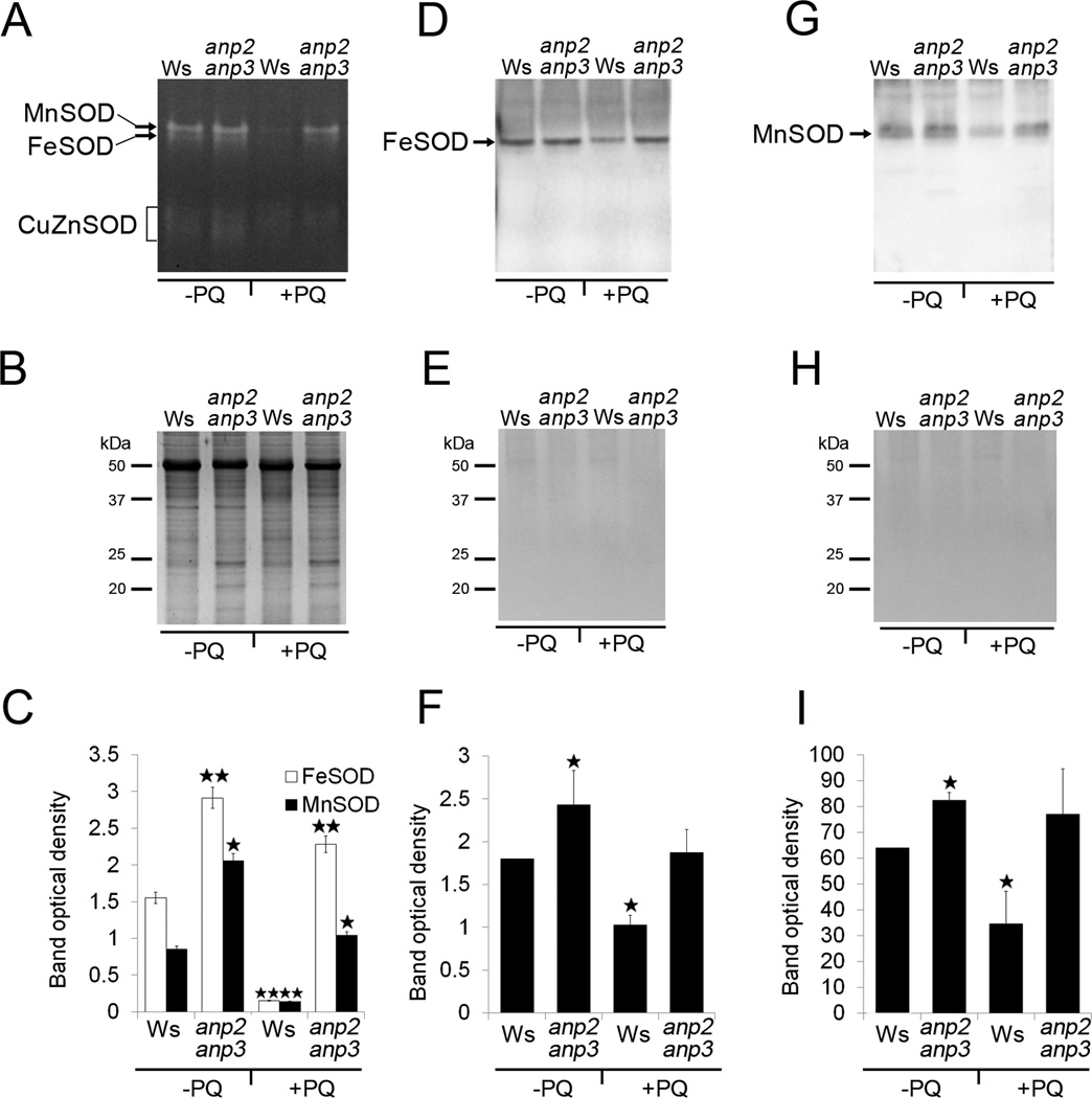
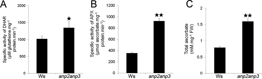
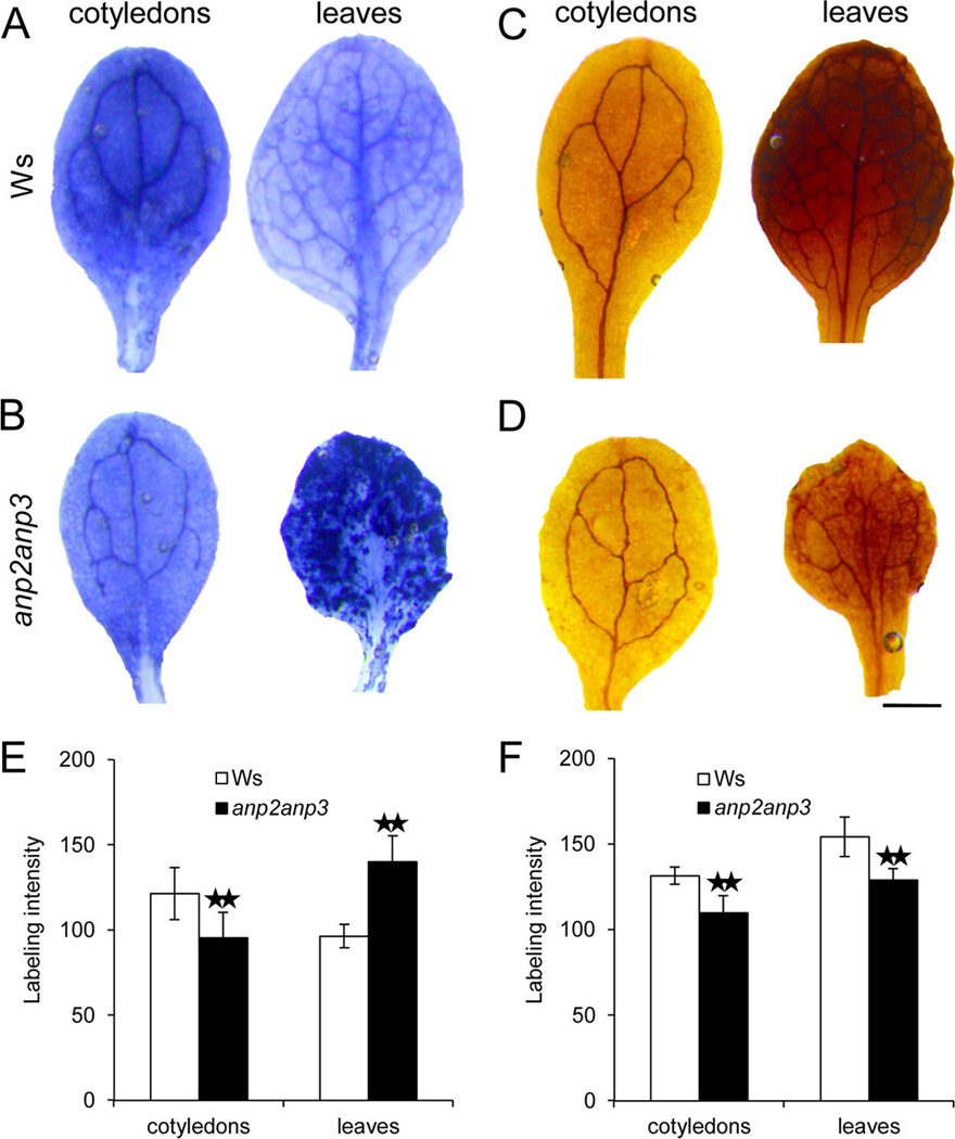
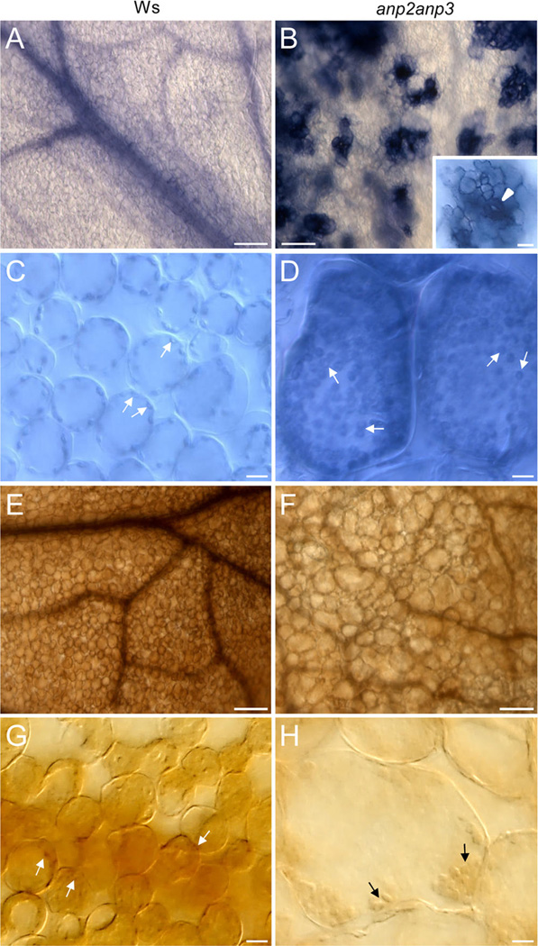
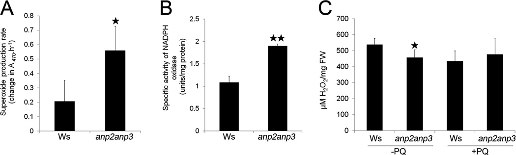
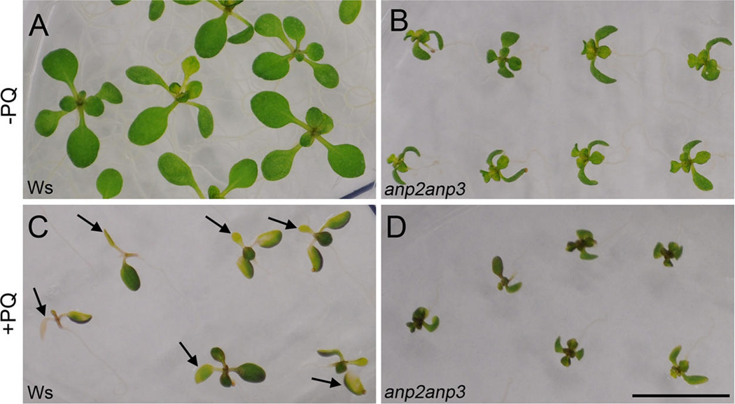
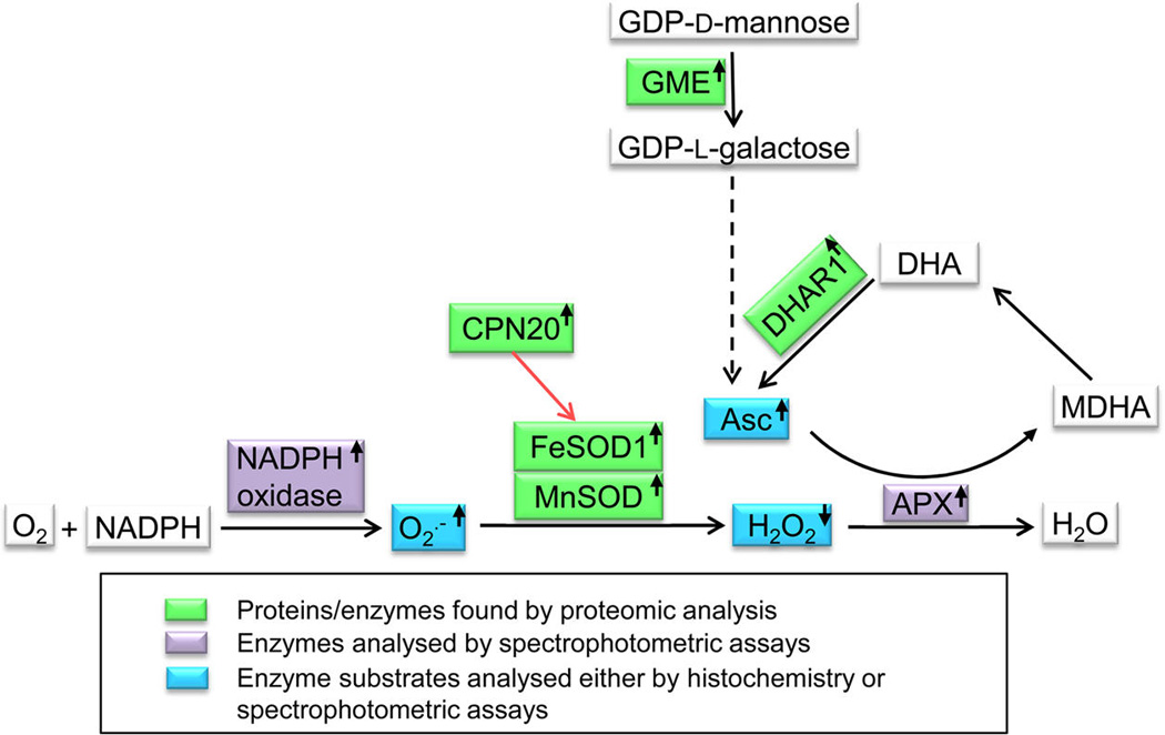
Disentanglement of functional complexity associated with plant mitogen-activated protein kinase (MAPK) signaling has benefited from transcriptomic, proteomic, phosphoproteomic, and genetic studies. Published transcriptomic analysis of a double homozygous recessive anp2anp3 mutant of two MAPK kinase kinase (MAPKKK) genes called Arabidopsis thaliana Homologues of Nucleus- and Phragmoplast-localized Kinase 2 (ANP2) and 3 (ANP3) showed the upregulation of stress-related genes. In this study, a comparative proteomic analysis of anp2anp3 mutant against its respective Wassilevskaja ecotype (Ws) wild type background is provided. Such differential proteomic analysis revealed overabundance of core enzymes such as FeSOD1, MnSOD, DHAR1, and FeSOD1-associated regulatory protein CPN20, which are involved in the detoxification of reactive oxygen species in the anp2anp3 mutant. The proteomic results were validated at the level of single protein abundance by Western blot analyses and by quantitative biochemical determination of antioxidant enzymatic activities. Finally, the functional network of proteins involved in antioxidant defense in the anp2anp3 mutant was physiologically linked with the increased resistance of mutant seedlings against paraquat treatment.

Keywords: ANP2; ANP3; Arabidopsis; antioxidant defense; mitogen-activated protein kinase kinase kinase; oxidative stress; proteomics; signaling.


Figures

Figure 1
Figure 1
Classification of proteins identified in the Arabidopsis anp2anp3 double mutant and Ws according to KEGG pathways.
Figure 2
Figure 2
Immunoblotting analysis of superoxide dismutase (SOD) isozymes in anp2anp3 mutant and wild type (Ws) seedlings. (A, B) Immunoblot detection (A) and band optical density quantification (B) of FeSOD1 (mean ± SD, N = 3); (C, D) Immunoblot detection (C) and band optical density quantification (D) of MnSOD (mean ± SD, N = 3). The right panels in A and C show Ponceau S staining of respective PVDF membranes. * in B, D indicates statistical significance of optical density difference at p < 0.05.
Figure 3
Figure 3
Isozyme pattern and immunoblotting on native PAGE gels of superoxide dismutase (SOD) in wild type (Ws) and anp2anp3 double mutant in control conditions (−PQ) and after paraquat treatment (+PQ). (A) SOD specific activity staining and (B) respective loading control represented by colloidal coomassie blue-stained SDS PAGE gels. Arrows in A indicate MnSOD and FeSOD, and the bracket shows CuZn SOD isozymes. (C) Quantification of the band densities in A (mean ± SD, N = 3). Asterisks indicate a statistically significant difference between Ws and anp2anp3 double mutant in control conditions as well as after paraquat treatment and between Ws in control conditions and after paraquat treatment as revealed by Student’s t test (* indicates statistical significance at p < 0.0S, ** indicates statistical significance at p < 0.01). (D and G) Immunoblots of FeSOD1 and MnSOD prepared on native PAGE gels using anti FeSOD1 and anti MnSOD antibodies. (E, H) Ponceau S staining of proteins (respective to D and G) transferred to PVDF membrane. (F, I) Quantification of the band densities in D and G (mean ± SD, N = 3). Asterisks indicate statistically significant differences in optical densities as revealed by Student’s t test (p < 0.05) between Ws and anp2anp3 double mutant in control conditions, as well as between Ws in control conditions and after paraquat treatment.
Figure 4
Figure 4
Quantitative demonstration of enzymatic activities within the ascorbate-glutathione cycle and ascorbate content in anp2anp3 double mutant. (A) Specific activity of dehydroascorbate reductase (DHAR; mean ± SD, N = 3). (B) Specific activity of ascorbate peroxidase (APX, mean ± SD, N = 3) and (C) total ascorbate content (mean ± SD, N = 3). * in A indicates statistical significance of enzymatic activities at p < 0.05, ** in B and C indicates statistical significance at p < 0.01.
Figure 5
Figure 5
Generation and distribution of superoxide (O2•−) and hydrogen peroxide (H2O2) in leaves and cotyledons of 10-day-old Arabidopsis plants of wild type Ws (A, C) and anp2anp3 double mutant (B, D). O2•− production was visualized as dark blue coloration by nitroblue tetrazolium (NBT) staining (A, B), and H2O2 production was visualized as dark brown coloration by 3,3′-diaminobenzidine (DAB) staining (C, D). Semiquantitative analysis of the intensity of NBT (E) and DAB (F) staining in cotyledons and leaves of wild type plants and anp2anp3 mutants. ** indicates statistical difference significant at a p value <0.01 as determined by Student’s t test (n = 34 for cotyledons and N = 76 for leaves in E, N = 30 for cotyledons and N = 58 for leaves in F). Bar represents 0.S mm for A–D.
Figure 6
Figure 6
Cellular pattern of superoxide (O2•−) and hydrogen peroxide (H2O2) distribution in leaves of wild type (Ws) plants (A, C, E, G) and anp2anp3 mutants (B, D, F, H). Nitroblue tetrazolium (NBT) staining revealed generation of O2•− in leaf vascular tissue and uniform distribution in leaf mesophyll cells in Ws plants (A), while in leaves of anp2anp3 mutant the staining showed a clustered pattern (B). Intensive staining in these isolated islands was localized to stomata (arrowhead), surrounding epidermal cells (inset in B), and mesophyll cells close to the stomatal cavity (B). In both Ws plants and anp2anp3 mutants, NBT staining in mesophyll cells was restricted mainly to chloroplasts (arrows in C, D). 3,3′-Diaminobenzidine (DAB) staining of Ws and anp2anp3 mutant leaves (E, F) revealed generation of H2O2 in vascular tissue and in mesophyll cells, which was more intensive in leaves of Ws plants (E). Staining in leaf mesophyll cells was localized mainly to chloroplasts (arrows in G, H). Bar = 100 µm (A, B, E, F), 20 µm (insert in B), and 10 µm(C, D, G, H).
Figure 7
Figure 7
Quantitative demonstration of superoxide (O2•−) production and H2O2 level in anp2anp3 double mutant. (A) Level of O2•− production in wild type (Ws) and anp2anp3 seedlings, presented as change in absorbance at 470 nm caused by O2•− -induced XTT reduction (mean ± SD, N = 3). (B) Specific activity of NADPH oxidase in anp2anp3 mutant and wild type seedlings (mean ± SD, N = 3). (C) H2O2 concentration in control (−PQ) and paraquat treated (+PQ) seedlings of Ws and anp2anp3. Asterisks indicate statistically significant difference between Ws and anp2anp3 double mutant (* indicates statistical significance at p < 0.0S, ** indicates statistical significance at p < 0.01).
Figure 8
Figure 8
Effect of oxidative stress on seedlings of anp2anp3 double mutant. (A, B) Arabidopsis seedlings of wild type (Ws) (A) and anp2anp3 mutant (B) grown under control conditions (−PQ). (C, D) Arabidopsis seedlings of Ws (C) and anp2anp3 double mutant (D) grown on 1/2 MS media supplemented with 0.S µM paraquat (+PQ). Three-day-old seedlings (wild type and mutant) were transferred to control and paraquat containing media and grown for 7 days. Note inhibition of the seedling growth and development as well as chlorophyll bleaching (arrows in C) in the leaves of Ws plants in comparison to the mutant plants. Bar represents 1 cm.
Figure 9
Figure 9
(A) Spatial distribution of the maximum quantum yield of photosystem II photochemistry (FV/FM) within Arabidopsis seedlings of wild type (Ws) and anp2anp3 double mutant obtained by chlorophyll fluorescence imaging. (B) Actual quantum yield of photosystem II electron transport (ΦPSII upper graph) and nonphotochemical chlorophyll fluorescence quenching (qN, bottom graph) during chlorophyll fluorescence induction in Arabidopsis seedlings of Ws (black, mean ± SD, N = 3) and anp2anp3 mutant (red, mean ± SD, N = 3).
Figure 10
Figure 10
Functional network of proteins/enzymes, reactive oxygen species, and redox-active compounds found by the current study to be modified in the anp2anp3 mutant (depicted in colored boxes). Red arrow shows activation, dashed arrow means metabolic pathway. The small arrows in boxes indicate upregulation or downregulation of individual components. Abbreviations: Asc = ascorbate, APX = ascorbate peroxidase, CPN20 = Chaperonin 20, DHAR 1 = dehydroascorbate reductase, GME = GDP-d-mannose 3′S′-epimerase, FeSOD = Fe-superoxide dismutase, MDHA = monodehydroascorbate, MnSOD = Mn-superoxide dismutase.

Similar articles

Cited by

References

    1. Rodriguez MCS, Petersen M, Mundy J. Mitogen-activated protein kinase signaling in plants. Annu. Rev. Plant Biol. 2010;61:621–649. - PubMed
    1. Šamajová O, Plíhal O, Al-Yousif M, Hirt H, Šamaj J. Improvement of stress tolerance in plants by genetic manipulation of mitogen-activated protein kinases. Biotechnol. Adv. 2013;31:118–128. - PubMed
    1. Andreasson E, Ellis B. Convergence and specificity in the Arabidopsis MAPK nexus. Trends Plant Sci. 2010;15:106–113. - PubMed
    1. Krysan PJ, Jester PJ, Gottwald JR, Sussman M. R An Arabidopsis mitogen-activated protein kinase kinase kinase gene family encodes essential positive regulators of cytokinesis. Plant Cell. 2002;14:1109–1120. - PMC - PubMed
    1. Sasabe M, Soyano T, Takahashi Y, Sonobe S, Igarashi H, Itoh TJ, Hidaka M, Machida Y. Phosphorylation of NtMAP6S–l by a MAP kinase down-regulates its activity of microtubule bundling and stimulates progression of cytokinesis of tobacco cells. Genes Dev. 2006;20:1004–1014. - PMC - PubMed

Publication types

MeSH terms