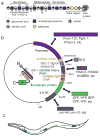
A transgenic approach to live imaging of heparan sulfate modification patterns
- PMID: 25325959
- PMCID: PMC5893304
- DOI: 10.1007/978-1-4939-1714-3_22
A transgenic approach to live imaging of heparan sulfate modification patterns
Abstract
Heparan sulfate (HS) glycosaminoglycan chains contain highly modified HS domains that are separated by sections of sparse or no modification. HS domains are central to the role of HS in protein binding and mediating protein-protein interactions in the extracellular matrix. Since HS domains are not genetically encoded, they are impossible to visualize and study with conventional methods in vivo. Here we describe a transgenic approach using previously described single chain variable fragment (scFv) antibodies that bind HS in vitro and on tissue sections with different specificities. By engineering a secretion signal and a fluorescent protein to the scFvs and transgenically expressing these fluorescently tagged antibodies in Caenorhabditis elegans, we are able to directly visualize specific HS domains in live animals (Attreed et al. Nat Methods 9(5):477-479, 2012). The approach allows concomitant colabeling of multiple epitopes, the study of HS dynamics and, could lend itself to a genetic analysis of HS domain biosynthesis or to visualize other nongenetically encoded or posttranslational modifications.
Figures


Similar articles
-
Imaging Glycosaminoglycan Modification Patterns In Vivo.Methods Mol Biol. 2022;2303:539-557. doi: 10.1007/978-1-0716-1398-6_42. Methods Mol Biol. 2022. PMID: 34626406
-
Conservation of anatomically restricted glycosaminoglycan structures in divergent nematode species.Glycobiology. 2016 Aug;26(8):862-870. doi: 10.1093/glycob/cww037. Epub 2016 Mar 13. Glycobiology. 2016. PMID: 26976619 Free PMC article.
-
Direct visualization of specifically modified extracellular glycans in living animals.Nat Methods. 2012 Apr 1;9(5):477-9. doi: 10.1038/nmeth.1945. Nat Methods. 2012. PMID: 22466794 Free PMC article.
-
Heparan Sulfate: Biosynthesis, Structure, and Function.Int Rev Cell Mol Biol. 2016;325:215-73. doi: 10.1016/bs.ircmb.2016.02.009. Epub 2016 Apr 13. Int Rev Cell Mol Biol. 2016. PMID: 27241222 Review.
-
Heparan sulfate-protein interactions--a concept for drug design?Thromb Haemost. 2007 Jul;98(1):109-15. Thromb Haemost. 2007. PMID: 17598000 Review.
Cited by
-
Imaging Glycosaminoglycan Modification Patterns In Vivo.Methods Mol Biol. 2022;2303:539-557. doi: 10.1007/978-1-0716-1398-6_42. Methods Mol Biol. 2022. PMID: 34626406
-
Conservation of anatomically restricted glycosaminoglycan structures in divergent nematode species.Glycobiology. 2016 Aug;26(8):862-870. doi: 10.1093/glycob/cww037. Epub 2016 Mar 13. Glycobiology. 2016. PMID: 26976619 Free PMC article.
-
HS, an Ancient Molecular Recognition and Information Storage Glycosaminoglycan, Equips HS-Proteoglycans with Diverse Matrix and Cell-Interactive Properties Operative in Tissue Development and Tissue Function in Health and Disease.Int J Mol Sci. 2023 Jan 6;24(2):1148. doi: 10.3390/ijms24021148. Int J Mol Sci. 2023. PMID: 36674659 Free PMC article. Review.
References
-
- Bernfield M, Götte M, Park PW, Reizes O, Fitzgerald ML, Lincecum J, Zako M. Functions of cell surface heparan sulfate proteoglycans. Annu Rev Biochem. 1999;68:729–777. - PubMed
-
- Bishop JR, Schuksz M, Esko JD. Heparan sulphate proteoglycans fine-tune mammalian physiology. Nature. 2007;446:1030–1037. - PubMed
-
- Bülow HE, Hobert O. The Molecular Diversity of Glycosaminoglycans Shapes Animal Development. Ann Rev Cell Dev Biol. 2006;22:375–407. - PubMed
Publication types
MeSH terms
Substances
Grants and funding
LinkOut - more resources
Full Text Sources
Other Literature Sources

