. 2014 Nov 10;42(20):12779-88.
doi: 10.1093/nar/gku951. Epub 2014 Oct 17.

Identification of a Mg2+-sensitive ORF in the 5'-leader of TRPM7 magnesium channel mRNA

Affiliations

Identification of a Mg2+-sensitive ORF in the 5'-leader of TRPM7 magnesium channel mRNA

Inna A Nikonorova et al. Nucleic Acids Res. .

Abstract

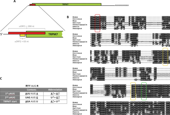
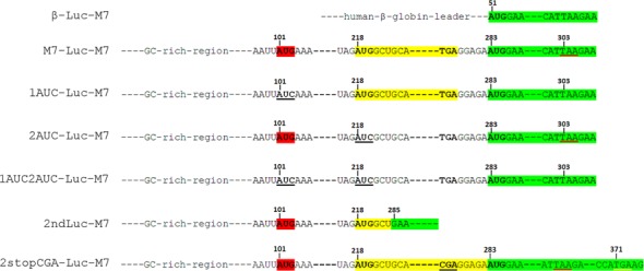
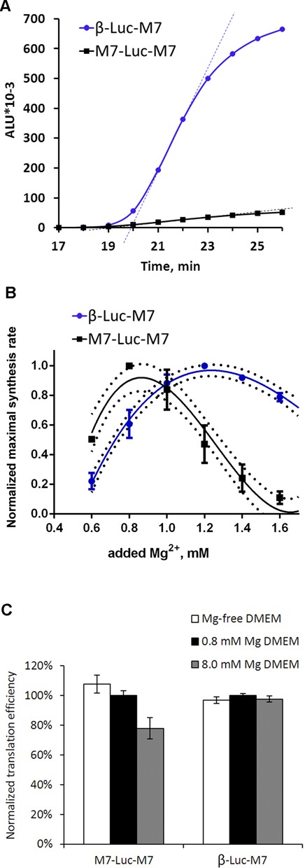
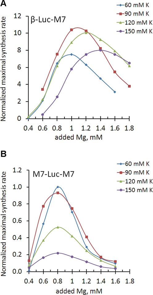
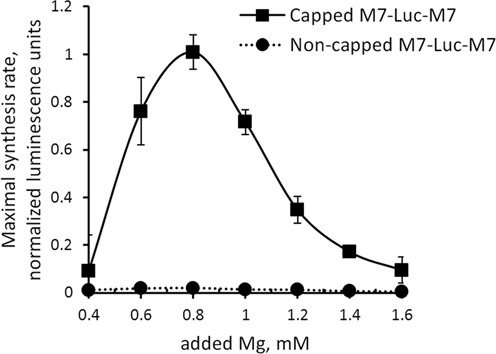
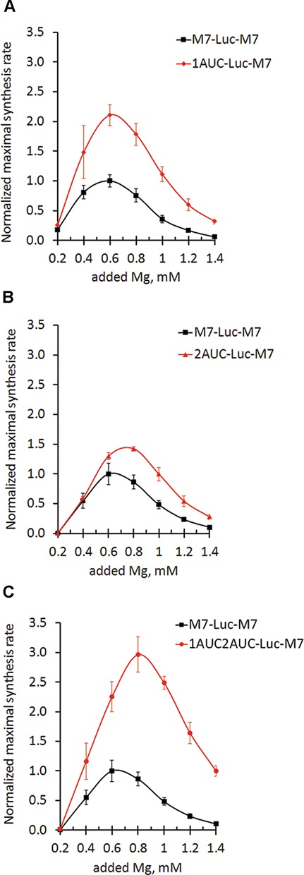
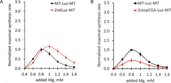
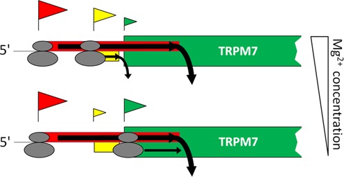
TRPM7 is an essential and ubiquitous channel-kinase regulating cellular influx of Mg2+. Although TRPM7 mRNA is highly abundant, very small amount of the protein is detected in cells, suggesting post-transcriptional regulation of trpm7 gene expression. We found that TRPM7 mRNA 5'-leader contains two evolutionarily conserved upstream open reading frames that act together to drastically inhibit translation of the TRPM7 reading frame at high magnesium levels and ensure its optimal translation at low magnesium levels, when the activity of the channel-kinase is most required. The study provides the first example of magnesium channel synthesis being controlled by Mg2+ in higher eukaryotes.


Figures

Figure 1.
Figure 1.
TRPM7 5′-leader contains two uORFs that are highly conserved across all vertebrates. (A) Schematic representation of TRPM7 mRNA organization with a close up on 5′-leader sequence. TRPM7 coding sequence is shown in green. The uORF1 (shown in red) overlaps the start codon of TRPM7 coding sequence. The uORF2 (shown in yellow) ends 5 nucleotides before the start codon of TRPM7 coding sequence. (B) Alignment of 5′ regions of TRPM7 mRNAs from different vertebrate species. The first uAUG (uAUG1) is marked in red, the second upstream AUG (uAUG2) and the corresponding termination codons are marked in yellow. The main AUG codon of TRPM7 coding sequence is marked in green. (C) Highly conserved contexts of the two uAUG codons and the main start codon of TRPM7 mRNA are indicated in the table. Nucleotides that match Kozak consensus for optimal initiation are underlined. Ideal Kozak consensus is presented above the table (R indicates purine nucleotide, Y indicates pyrimidine nucleotide).
Figure 2.
Figure 2.
Constructs designed to test the ability of TRPM7 5′-leader to direct translation of a luciferase reporter sequence. The main reading frame, coding for Firefly luciferase, is marked in green, the uORF2 is marked in yellow, and the AUG codon of the uORF1 is marked in red. Stop codon that terminates translation of the uORF1 is underlined in red. Mutations of TRPM7 5′-leader are underlined in black. Correspondence of a few key nucleotides of TRPM7 5′-leader mutant forms to positions of the same nucleotides in M7-Luc-M7 construct is indicated with numbers. The random stop codon that terminates translation initiated by the second uAUG in 2stopCGA-Luc-M7 construct is underlined in yellow.
Figure 3.
Figure 3.
Inefficient translation directed by TRPM7 5′-leader can be activated by lowering Mg2+concentration. (A) Kinetic curves of translation reactions directed by β-globin (circles) and TRPM7 (squares) 5′-leaders conducted under conditions of 1 mM of added magnesium acetate and 120 mM of added potassium acetate. Dashed lines show the maximal slopes of the kinetic curves, which reflect maximal synthesis rates of translation reactions. (B) Dependence of the maximal protein synthesis rates on Mg2+ concentration is given for translation reactions carried out at 120 mM of added K+. For each construct on diagram B, the maximal synthesis rate at each Mg2+ concentration was normalized to the value at the optimal Mg2+ concentration. Error bars represent standard deviation (SD) of three experiments. (C) Sensitivity of TRPM7 leader-directed translation measured in vivo to Mg2+ concentration in the medium. Cells were transfected with either β-Luc-M7 or M7-Luc-M7 mRNAs under different culturing conditions (DMEM containing either 0 mM, 0.8 mM or 8.0 mM of magnesium). Columns represent Firefly luciferase activity measured 3 h post transfection and normalized to the value of Renilla luciferase that was produced from an mRNA cotransfected with the constructs of interest. Error bars represent SD of two independent experiments performed in duplicates.
Figure 4.
Figure 4.
Magnesium optimum of translation directed by TRPM5 5′-leader is not perturbed by varying potassium concentration. Dependence of maximal synthesis rate on magnesium concentration at different potassium levels (diamonds—60 mM, squares—90 mM, triangles—120 mM and circles—150 mM of added potassium acetate). Panel (A) represents translation directed by β-globin 5′-leader. Panel (B) represents translation directed by TRPM7 5′-leader. Maximal synthesis rates are normalized to the value of M7-Luc-M7 translation at 60 mM of added potassium at the optimal magnesium concentration (0.8 mM of added magnesium acetate). All the reactions used to reconstruct magnesium dependencies on this figure were performed using the same lysate preparation.
Figure 5.
Figure 5.
TRPM7 5′-leader utilizes 5′-cap-dependent mechanism of translation initiation. Translation directed by a non-capped construct is inefficient as compared to the capped M7-Luc-M7 construct.
Figure 6.
Figure 6.
TRPM7 uORF1 and uORF2 reveal different roles in translation of the main coding sequence. Different dependences of translation efficiency on magnesium concentrations: squares represent maximal synthesis rates of translation reactions directed by TRPM7 5′-leader. Translation reactions directed by TRPM7 5′-leader mutant forms are indicated by diamonds for 1AUC-Luc-M7 (A), triangles for 2AUC-Luc-M7 (B) and circles for 1AUC2AUC-Luc-M7 (C). Maximal synthesis rates are normalized to the value of M7-Luc-M7 translation at the optimal magnesium concentration. Error bars represent SD of at least three experiments.
Figure 7.
Figure 7.
(A) TRPM7 uORF2 has higher magnesium optimum of translation initiation. Graph shows dependences of translation rates of uORF2 (circles) and the main coding sequence (squares) on magnesium concentration. (B) Stop codon of uORF2 contributes to the initiation of translation of the main reading frame. Graph shows translation rates of 2stopCGA-Luc-M7 (triangles) and M7-Luc-M7 (squares) mRNAs. Maximal synthesis rates were normalized to the value of M7-Luc-M7 translation at the optimal magnesium concentration. Error bars represent SD of at least three experiments.
Figure 8.
Figure 8.
Proposed model of regulation of TRPM7 protein synthesis based on scanning mechanism of translation initiation. Flag above an AUG codon represents an initiation event on that AUG. The size of the flag reflects probability of initiation on the corresponding AUG codon. Most of the time translation is initiated at the first uAUG codon so that the uORF1, which overlaps the start codon of TRPM7 protein, gets translated, preventing synthesis of TRPM7 protein. In a small number of cases 40S scanning subunits are able to bypass the uAUG1 and continue scanning. Under conditions of high magnesium, scanning ribosomal subunits are more likely to initiate at the uAUG2 (upper scenario) and translate the uORF2, reducing the amount of ribosomes reaching the main start codon. Under conditions of low magnesium, scanning 40S subunits are more likely to bypass the uAUG2 and to reach the start codon of TRPM7 coding sequence (lower scenario).

Similar articles

Cited by

References

    1. Fujiwara Y., Minor D.L. X-ray crystal structure of a TRPM assembly domain reveals an antiparallel four-stranded coiled-coil. J. Mol. Biol. 2008;383:854–870. - PMC - PubMed
    1. Nadler M.J., Hermosura M.C., Inabe K., Perraud A.L., Zhu Q., Stokes A.J., Kurosaki T., Kinet J.P., Penner R., Scharenberg A.M., et al. LTRPC7 is a Mg·ATP-regulated divalent cation channel required for cell viability. Nature. 2001;411:590–595. - PubMed
    1. Demeuse P., Penner R., Fleig A. TRPM7 channel is regulated by magnesium nucleotides via its kinase domain. J. Gen. Physiol. 2006;127:421–434. - PMC - PubMed
    1. Deason-Towne F., Perraud A-L, Schmitz C. The Mg2+ transporter MagT1 partially rescues cell growth and Mg2+ uptake in cells lacking the channel-kinase TRPM7. FEBS Lett. 2011;585:2275–2278. - PMC - PubMed
    1. Sahni J., Song Y., Scharenberg A.M. The B. subtilis MgtE magnesium transporter can functionally compensate TRPM7-deficiency in vertebrate B-cells. PloS One. 2012;7:e44452. - PMC - PubMed

Publication types