. 2014 Dec 26;289(52):36166-78.
doi: 10.1074/jbc.M114.612630. Epub 2014 Oct 17.

Identification of a novel recycling sequence in the C-tail of FPR2/ALX receptor: association with cell protection from apoptosis

Affiliations

Identification of a novel recycling sequence in the C-tail of FPR2/ALX receptor: association with cell protection from apoptosis

Dawn Thompson et al. J Biol Chem. .

Abstract

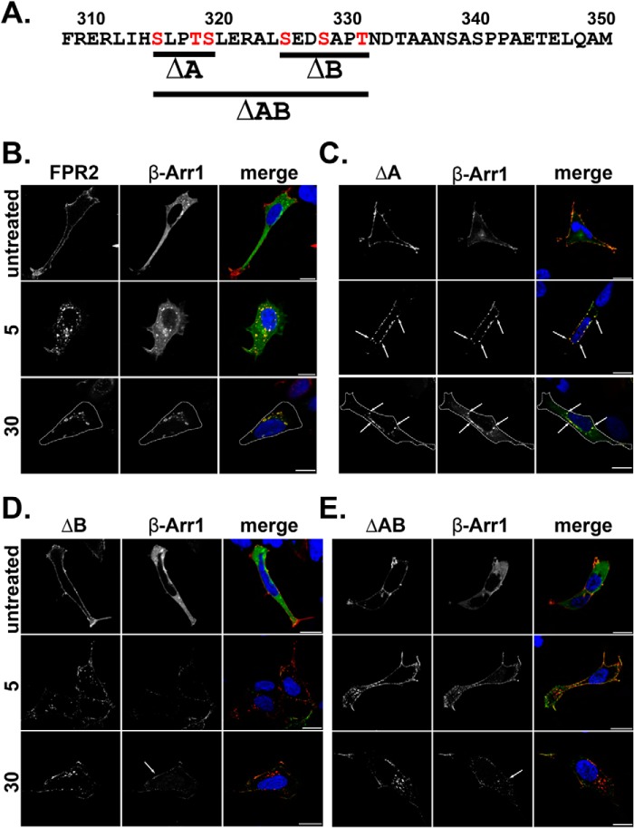
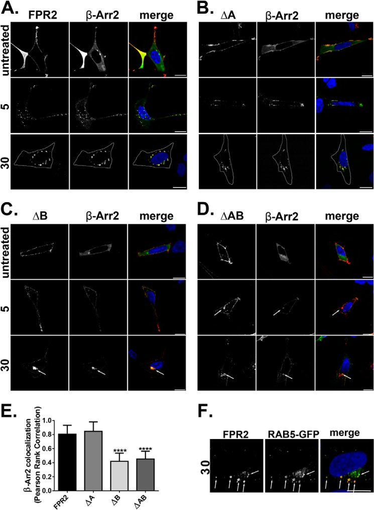
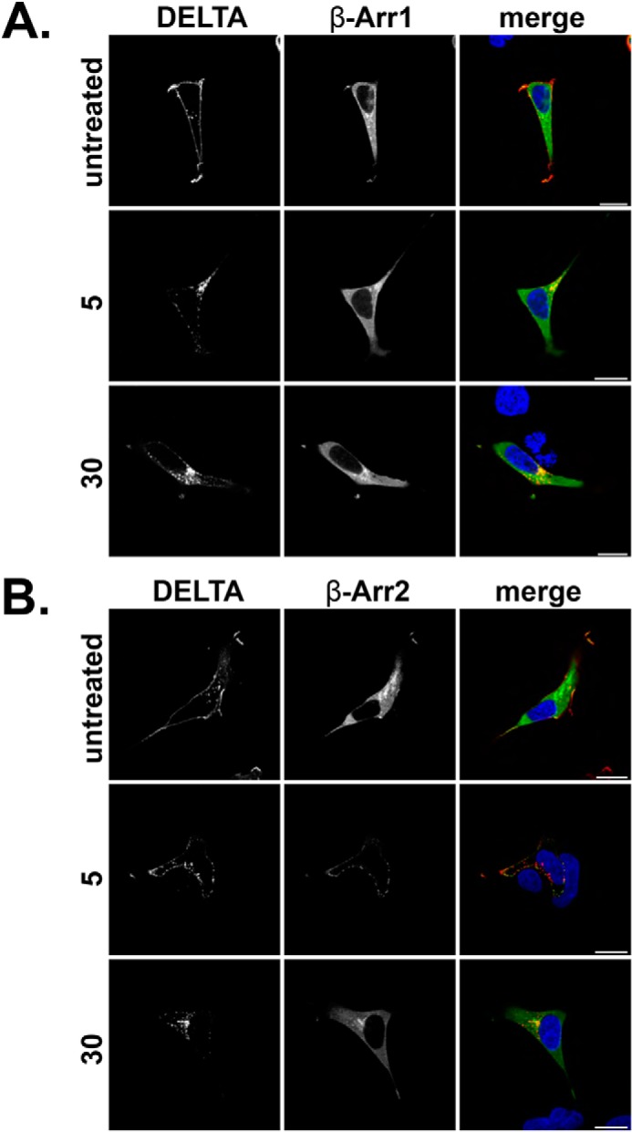
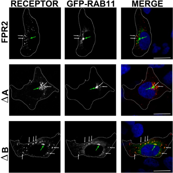
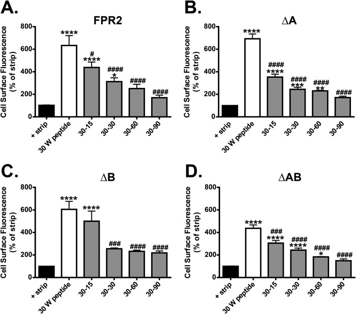
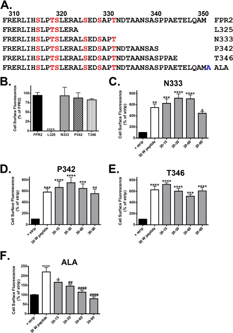
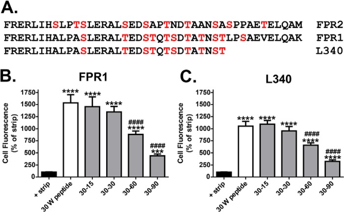
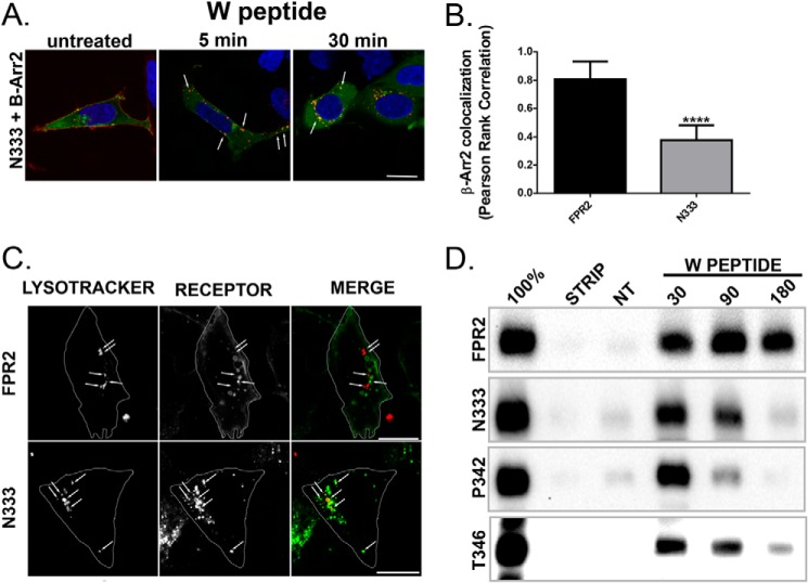
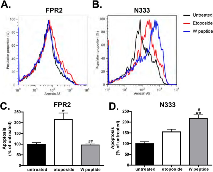
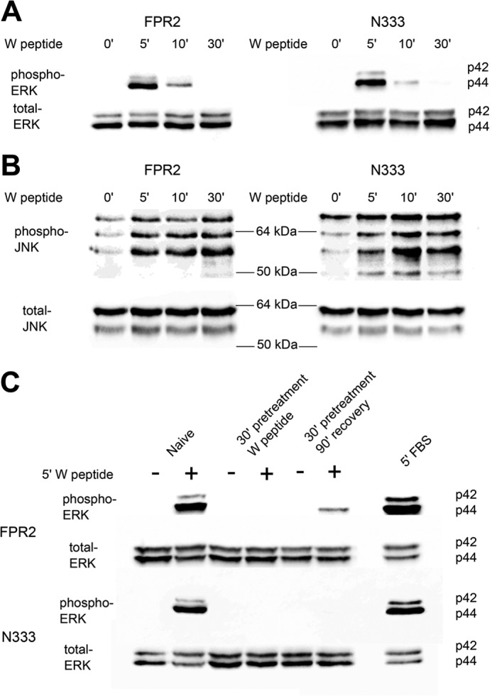
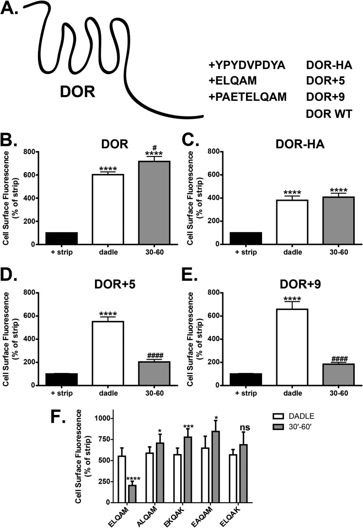
Formyl-peptide receptor type 2 (FPR2; also called ALX because it is the receptor for lipoxin A4) sustains a variety of biological responses relevant to the development and control of inflammation, yet the cellular regulation of this G-protein-coupled receptor remains unexplored. Here we report that, in response to peptide agonist activation, FPR2/ALX undergoes β-arrestin-mediated endocytosis followed by rapid recycling to the plasma membrane. We identify a transplantable recycling sequence that is both necessary and sufficient for efficient receptor recycling. Furthermore, removal of this C-terminal recycling sequence alters the endocytic fate of FPR2/ALX and evokes pro-apoptotic effects in response to agonist activation. This study demonstrates the importance of endocytic recycling in the anti-apoptotic properties of FPR2/ALX and identifies the molecular determinant required for modulation of this process fundamental for the control of inflammation.

Keywords: Apoptosis; Arrestin; Cell Sorting; Endocytosis; G-protein-coupled Receptor (GPCR); Receptor Recycling.


Figures

FIGURE 1.
FIGURE 1.
Mutation of serines and threonines in the C-tail of FPR2 changes β-Arr1 binding patterns. A, illustration of constructed phosphorylation mutants, ΔA, ΔB, and ΔAB, where red amino acids were mutated to alanines. B–E, HEK293 cells expressing N-terminally FLAG-tagged receptor constructs (FPR2, ΔA, ΔB, or ΔAB) and EGFP-tagged β-Arr1. Cells were fed with anti-FLAG M1 antibody to label receptor and treated as indicated (500 nm W peptide), then fixed, permeabilized, and incubated with secondary antibody prior to confocal microscopy. Representative images are shown with scale bars equal to 20 μm, and dotted lines mark the cell boundary.
FIGURE 2.
FIGURE 2.
Mutation of serines and threonines in the C-tail of FPR2 changes β-Arr2 binding patterns. A–D, HEK293 cells expressing N-terminally FLAG-tagged receptor constructs (FPR2, ΔA, ΔB or ΔAB) and EGFP-tagged β-Arr2. Cells were fed with anti-FLAG M1 antibody to label receptor and treated as indicated (500 nm W peptide) and then fixed, permeabilized, and incubated with secondary antibody and visualized using confocal microscopy. E, quantification of β-Arr2 association with receptor constructs at 30 min of W peptide treatment expressed as Pearson's correlation coefficient. Data are represented as the mean co-localization of at least 30 cells performed on three separate occasions and analyzed using one-way ANOVA with Bonferroni's t test where ****, p ≤ 0.001 when compared with WT FPR2. Error bars indicate mean ± S.D. F, cells co-expressing WT N-terminally FLAG-tagged FPR2 and EGFP Rab5 were fed with M1 antibody as in A and incubated for 20 min with W peptide. Representative images are shown with scale bars equal to 20 μm. Cell boundary is marked by a dotted line.
FIGURE 3.
FIGURE 3.
DOR recruits β-Arr2. A and B, HEK293 cells were transfected with N-terminally FLAG-tagged DOR and either EGFP β-Arr1 (A) or EGFP β-Arr2 (B) and fed with M1 antibody (1:1000) to label mature cell surface receptors. Cells were untreated or stimulated with DADLE (10 μm) for 5 or 30 min, fixed, permeabilized, and incubated with secondary antibody (anti-mouse IgG2b Alexa Fluor® 594, 1:1000) and visualized by confocal microscopy. Representative images are shown with scale bars equal to 20 μm. DELTA, δ-opioid receptor.
FIGURE 4.
FIGURE 4.
Mutation of the ΔB residues confers retention in the Rab11 compartment. Cells expressing WT N-terminally FLAG-tagged FPR2, ΔA, or ΔB were co-expressed with EGFP Rab11 and fed with M1 antibody as in Figs. 1 and 2 and incubated for 30 min with W peptide. Cells were then fixed, permeabilized, and incubated with secondary antibody (anti-mouse IgG2b Alexa Fluor ® 594, 1:1000) and visualized using confocal microscopy. Representative images are shown with scale bars equal to 20 μm (green arrow indicates the Rab11 recycling compartment, and white arrows show co-localization).
FIGURE 5.
FIGURE 5.
Mutation of the serines and threonines in the C-tail of FPR2 has no effect on receptor recycling. A–D, flow cytometry was used to analyze the endocytosis and recycling properties of FPR2, ΔA, ΔB, and ΔAB (see “Experimental Procedures”). HEK293 cells expressing either WT or mutant receptors were labeled with M1-conjugated Alexa Fluor® 647, stimulated with W peptide (500 nm) for 30 min, and either washed in PBS or returned to the incubator in the presence of PBS/EDTA for 15, 30, 60, or 90 min to monitor the degree of recycling. Data are represented as the mean of at least four independent experiments performed in duplicate analyzed using one-way ANOVA with Bonferroni's t test where ****, p ≤ 0.0001, ***, p ≤ 0.001, **, p ≤ 0.01, *, p ≤ 0.05 when compared with untreated controls or ####, p ≤ 0.0001, ###, p ≤ 0.001, #, p ≤ 0.05 when compared with W peptide-treated samples. Error bars indicate mean ± S.D.
FIGURE 6.
FIGURE 6.
Truncation of the FPR2 C-tail prevents receptor recycling. A, illustration of truncation mutants where stop codons were introduced at amino acids Leu-325, Asn-333, Pro-342, or Thr-346 or the addition of a single alanine to the end of the C-tail (ALA). B, WT FPR2 or the truncation mutants were assessed for cell surface expression by flow cytometry. C–F, the endocytosis and recycling properties of N333-stop (N333), P342-stop (P342), T346-stop (T346), and ALA was analyzed by flow cytometry as in Fig. 5. Data are represented as the mean of at least four independent experiments performed in duplicate analyzed using one-way ANOVA with Bonferroni's t test where ****, p ≤ 0.0001, ***, p ≤ 0.001, **, p ≤ 0.01, *, p ≤ 0.05 when compared with untreated controls or ##, p ≤ 0.01, ####, p ≤ 0.0001 when compared with W peptide-treated samples. There were no significant differences in samples when compared with W peptide-treated samples. Error bars indicate mean ± S.D.
FIGURE 7.
FIGURE 7.
Truncation of the FPR1 C-tail does not prevent receptor recycling. A, alignment of FPR1 and FPR2 C-tail sequences and illustration of truncation mutant where a stop codon was introduced at amino acid Leu-340 in the FPR1 backbone. L340, L340-stop. B and C, the endocytosis and recycling properties of WT FPR1 and Leu-340 were analyzed by flow cytometry as in Fig. 5. Data are represented as the mean of at least four independent experiments performed in duplicate analyzed using one-way ANOVA with Bonferroni's t test where ****, p ≤ 0.0001, ***, p ≤ 0.001 when compared with untreated controls or ####, p ≤ 0.0001 when compared with W peptide-treated samples. Error bars indicate mean ± S.D.
FIGURE 8.
FIGURE 8.
Attenuated recycling leads to lysosomal targeting and receptor down-regulation. A, HEK293 cells expressing N-terminally FLAG-tagged N333-stop and EGFP-tagged β-Arr2 were fed with anti-FLAG M1 antibody as in Fig. 2, treated with W peptide, and then fixed, permeabilized, and incubated with secondary antibody and visualized using confocal microscopy. Representative images are shown with scale bars equal to 20 μm, and arrows indicate examples of co-localization. B, quantification of β-Arr2 association with FPR2 and N333-stop (N333) at 30-min W peptide treatment expressed as Pearson's correlation. Data are represented as the mean co-localization of at least 30 cells performed on three separate occasions and analyzed using one-way ANOVA with Bonferroni's t test where ****, p ≤ 0.001 when compared with WT FPR2. Error bars indicate mean ± S.D. C, HEK293 cells stably expressing FPR2 or N333-stop were labeled with anti-FLAG M1 antibody and LysoTracker and incubated for 90 min with 500 nm W peptide. Representative confocal images are shown with scale bars equal to 20 μm, and arrows indicate the lysosomal compartment. D, HEK293 cells stably expressing FPR2, N333-stop (N333), P342-stop (P343), or T346-stop (T346) were surface-biotinylated and either untreated or stimulated with 500 nm W peptide for 30, 90, or 180 min. Receptor fate was assessed after immunoprecipitation with anti-FLAG M2 antibody, subsequent separation by SDS-PAGE electrophoresis, and streptavidin overlay. The 100% lane shows total surface receptor labeling, and the STRIP lane indicates the efficiency of the biotin cleavage. NT, non-treatment. Representative immunoblots are shown.
FIGURE 9.
FIGURE 9.
Attenuated recycling leads to cellular apoptosis. A and B, cells expressing either FPR2 (A) or N333-stop (N333) (B) were untreated or treated for 5 h with either etoposide or W peptide, stained with propidium iodide and annexin V, and analyzed using flow cytometry. A representative trace is shown. C and D, quantification of three experiments performed in duplicate and analyzed using one-way ANOVA with Bonferroni's t-tests. **, p ≤ 0.01, *, p ≤ 0.05 when compared with untreated controls. ##, p ≤ 0.01 or #, p ≤ 0.05 when compared with W peptide-treated samples. Error bars indicate mean ± S.D.
FIGURE 10.
FIGURE 10.
MAPK signaling of FPR2 and N333-stop. A and B, HEK293 cells stably expressing either N-terminally FLAG-tagged WT FPR2 or N333-stop (N333) were serum-starved 4 h prior to experimentation. Cells were untreated or stimulated with 500 nm W peptide for 5, 10, and 30 min, lysed, separated by SDS-PAGE, and probed for phospho-ERK 1/2 (A) or JNK (B), and then stripped and reprobed for total ERK 1/2 and JNK. C, for resensitization, phospho-ERK 1/2 was investigated where cells were either untreated, stimulated for 5 min, pretreated with agonist for 30 min, and then re-challenged with vehicle or drug (desensitized) or pretreated for 30 min, washed, and allowed to recover for 90 min at 37 °C before a final re-challenge for 5 min with agonist or vehicle (resensitization and recycling). Representative blots are shown of at least three independent experiments.
FIGURE 11.
FIGURE 11.
FPR2 contains a specific motif that is able to facilitate recycling of the degrading δ-opioid receptor. A, chimeras of DOR as introduced by PCR: DOR-HA (YPYDVPDYA), DOR+5 (ELQAM), and DOR+9 (PAETELQAM). B–E, cells transiently expressing WT or chimeric constructs were assessed for endocytosis and recycling by flow cytometry (see “Experimental Procedures”). Cells were labeled as in Fig. 5 and either untreated or stimulated for 30 min with DADLE (10 μm) and then washed with PBS/EDTA to assess acute endocytosis, or stimulated and returned to the incubator for 60 min in the presence of PBS/EDTA to assess receptor recycling. F, point mutations were introduced to the DOR+5 backbone to elucidate specific amino acids within this motif critical for receptor recycling. Data are represented as the mean of at three independent experiments performed in duplicate analyzed using one-way ANOVA with Bonferroni's t test where ****, p ≤ 0.0001, ***, **, p ≤ 0.01, *, p ≤ 0.05 when compared with untreated controls or ####, p ≤ 0.0001 or #, p ≤ 0.05 when compared with DADLE-treated samples. Error bars indicate mean ± S.D.

Similar articles

Cited by

References

    1. Nathan C., Ding A. (2010) Nonresolving inflammation. Cell 140, 871–882 - PubMed
    1. Babbin B. A., Laukoetter M. G., Nava P., Koch S., Lee W. Y., Capaldo C. T., Peatman E., Severson E. A., Flower R. J., Perretti M., Parkos C. A., Nusrat A. (2008) Annexin A1 regulates intestinal mucosal injury, inflammation, and repair. J. Immunol. 181, 5035–5044 - PMC - PubMed
    1. Levy B. D., Vachier I., Serhan C. N. (2012) Resolution of inflammation in asthma. Clin. Chest Med. 33, 559–570 - PMC - PubMed
    1. Hanyaloglu A. C., von Zastrow M. (2008) Regulation of GPCRs by endocytic membrane trafficking and its potential implications. Annu. Rev. Pharmacol. Toxicol. 48, 537–568 - PubMed
    1. Dufton N., Perretti M. (2010) Therapeutic anti-inflammatory potential of formyl-peptide receptor agonists. Pharmacol. Ther. 127, 175–188 - PubMed

Publication types

LinkOut - more resources