. 2014 Dec 5;289(49):34267-83.
doi: 10.1074/jbc.M114.598201. Epub 2014 Oct 16.

Structure-function analyses of human kallikrein-related peptidase 2 establish the 99-loop as master regulator of activity

Affiliations

Structure-function analyses of human kallikrein-related peptidase 2 establish the 99-loop as master regulator of activity

Wolfgang Skala et al. J Biol Chem. .

Abstract

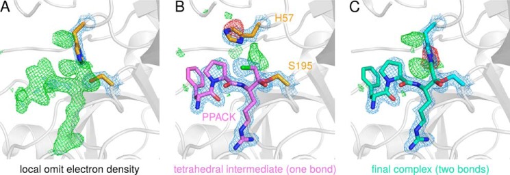
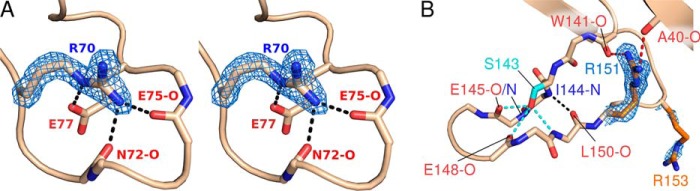
Human kallikrein-related peptidase 2 (KLK2) is a tryptic serine protease predominantly expressed in prostatic tissue and secreted into prostatic fluid, a major component of seminal fluid. Most likely it activates and complements chymotryptic KLK3 (prostate-specific antigen) in cleaving seminal clotting proteins, resulting in sperm liquefaction. KLK2 belongs to the "classical" KLKs 1-3, which share an extended 99- or kallikrein loop near their non-primed substrate binding site. Here, we report the 1.9 Å crystal structures of two KLK2-small molecule inhibitor complexes. In both structures discontinuous electron density for the 99-loop indicates that this loop is largely disordered. We provide evidence that the 99-loop is responsible for two biochemical peculiarities of KLK2, i.e. reversible inhibition by micromolar Zn(2+) concentrations and permanent inactivation by autocatalytic cleavage. Indeed, several 99-loop mutants of KLK2 displayed an altered susceptibility to Zn(2+), which located the Zn(2+) binding site at the 99-loop/active site interface. In addition, we identified an autolysis site between residues 95e and 95f in the 99-loop, whose elimination prevented the mature enzyme from limited autolysis and irreversible inactivation. An exhaustive comparison of KLK2 with related structures revealed that in the KLK family the 99-, 148-, and 220-loop exist in open and closed conformations, allowing or preventing substrate access, which extends the concept of conformational selection in trypsin-related proteases. Taken together, our novel biochemical and structural data on KLK2 identify its 99-loop as a key player in activity regulation.

Keywords: 99-Loop; Autolytic Cleavage; Conformational Selection; Crystallography; Enzyme Kinetics; Kallikrein; Prostate Cancer; Serine Protease; Substrate Specificity; Zinc Inhibition.


Figures

FIGURE 1.
FIGURE 1.
Purification scheme for recombinant KLK2. Purification comprised negative IEC, activation of pro-KLK2 by EK, immobilized metal ion affinity chromatography (IMAC), BENAC, and size exclusion chromatography (SEC). Success of the individual steps was routinely monitored by SDS-PAGE. +DTT and −DTT indicates whether gels were run under reducing or oxidizing conditions, respectively. M, marker; L, load; F, flow-through; W, wash; E, elution; AU, absorbance units.
FIGURE 2.
FIGURE 2.
KLK2 kinetics and substrate specificity. a–c, all measurements involved 150 nm wild type KLK2 and 250 μm Bz-PFR-pNA. The pH optimum was determined in 100 mm SPG buffer. Relative velocities vrel are relative to the highest velocity in the respective panel. d, substrate specificity of KLK2 as determined by a diverse positional scanning library (30). The y axis represents the substrate cleavage rate relative to the highest rate observed for this position. The x axis indicates the amino acid held constant at each position (n, norleucine).
FIGURE 3.
FIGURE 3.
Primary, secondary, and tertiary structure of KLK2. A, overall structure of KLK2 in complex with benzamidine (cyan), shown in standard orientation (79), in which the substrates bind to non-primed subsites from the left and to primed subsites to the right of the scissile bond. Helices (blue), β-strands (red), and the 75-, 99-, 148-, and 220-loop (green) are shown as ribbons. Disulfide bridges (yellow), the active site residues Ser-214, Asp-102, His-57, and Ser-195 (orange), and the stabilizing salt bridge Ile-16–Asp-194 and Asp-189 at the bottom of the S1 pocket (gray) are drawn as sticks. B, molecular surface of KLK2 bound to PPACK. Colors relate to the electrostatic surface potential (−10 to +10 kBT), which was calculated by APBS (104). Note the negatively charged (red) substrate binding site at the front of the molecule and the positively charged (blue) surface next to the C-terminal α-helix at the back. C, structure-based amino acid sequence alignment of KLKs 1–5, bovine trypsin, and bovine chymotrypsin A. Residue numbers according to the chymotrypsinogen numbering scheme appear on top. The secondary structure of KLK2 is indicated below each row. A light or dark gray background denotes residues that are similar in at least four sequences or completely conserved, respectively. The remaining colors correspond to A. Minor deletions with respect to chymotrypsin A (bCTRA) are found in surface loops after His-36 and Lys-61, Thr-125, and Asn-202 as well as minor insertions after Trp-186, Pro-223, and the C-terminal Asn-245. The 11-residue insertion following Asn-95 is known as extended 99- or kallikrein loop of the classical KLKs 1–3. The alignment was prepared with STRAP (62) and visualized with TeXshade (71).
FIGURE 4.
FIGURE 4.
Different KLK2-PPACK complexes that form during the inhibition reaction. A, positive difference electron density (green), which was generated by refining KLK2-PPACK data in the absence of the ligand, indicates missing atoms in the substrate binding site. B, model of the tetrahedral intermediate of the inhibition reaction. This intermediate forms one covalent bond to Ser-195 (0G6-C2 to Ser-195-Oγ); the chlorine atom of the chloromethyl ketone group is still present. The tetrahedral intermediate is present in the crystal with low occupancy. C, the final KLK2-PPACK complex, which is characterized by a second covalent bond between the inhibitor and its target (0G6-C3 to His-57-Nϵ2). Note that the deposited KLK2-PPACK coordinates (PDB ID 4nff) model the latter situation only. Maximum likelihood-based difference electron densities are drawn in blue (2mFobsDFcalc map contoured at 1.5σ), green, and red (mFobsDFcalc map contoured at +3σ and -3σ, respectively). Structure factors and electron densities were calculated by REFMAC (37) and fft (67), respectively. Because crystallographic programs do not contain parameters for the two covalent bonds between PPACK and KLK2, they were generated with the software JLigand (39) and written to a crystallographic information file (CIF), which was employed in crystallographic refinement (supplemental File S1).
FIGURE 5.
FIGURE 5.
KLK2 surface loops. A, stereo view of the 75-loop. The guanidinium group of Arg-70 interacts with putative calcium ligands, thereby precluding calcium binding. Hydrogen bonds are shown as black, dashed lines. B, the 148-loop is stabilized by hydrogen bonds that involve Ser-143 (cyan, dashed lines), Arg-151 (red, dashed lines), or backbone atoms only (black, dashed line). Maximum-likelihood 2mFobsDFcalc electron density contoured at 1.5σ is shown in blue for the side chains of arginines 70, 151, and 153.
FIGURE 6.
FIGURE 6.
99-loops of the KLK family. A, an alignment of all KLK 99-loops whose structures are known generally classifies them as either short/intermediate (red) or long (blue, semitransparent). B, secondary structure of the long KLK3 99-loop, colored according to C. C, in the structure-based alignment of 99-loop sequences (right), each residue is labeled by the DSSP single-letter code for its secondary structure (instead of the single-letter amino acid code): B, isolated β-bridge; E, extended β-sheet; G, 310-helix; S, bend; T, turn. In addition, M denotes missing residues, dashed lines indicate gaps, and continuous lines represent residues that lack secondary structure. Sequence numbers appear on top, and brackets below the alignment label conserved secondary structure elements. After structural superposition, backbone Cα distances, i.e. pairwise r.m.s.d. values, for the corresponding 99-loop stretches were calculated with a script in the molecular graphics program PyMOL (supplemental File S2), resulting in a distance matrix. The UPGMA dendrogram (left) is based upon pairwise 99-loop Cα r.m.s.d. and organizes the 99-loops in six clusters, which are indicated to the right of the alignment. PDB codes are given in brackets. The dendrogram was calculated by PHYLIP (64) and drawn by TreeGraph 2 (72). D, representative members of the six 99-loop clusters I-V and V*. The double-headed arrow indicates that cluster V and V* loops probably represent interconvertible open and closed conformations.
FIGURE 7.
FIGURE 7.
KLK2 inactivation and inhibition kinetics. a, time-dependent activity loss points toward a second-order reaction mechanism. The regression curve illustrates the corresponding rate law, d[KLK2]/dt = −2 k [KLK2]2, with [KLK2]0 = 100% and k = 0.014 ± 0.002 d−1, resulting in a time dependence of 1/(2kt + 1). Inset, time-dependent 99-loop cleavage as evident from nonreducing SDS-PAGE. F, full-length KLK2; C, C-terminal fragment (His-95f–Pro-245a); N, N-terminal fragment (Ile-16–Lys-95e). Shown are Lineweaver-Burk (b) and Eadie-Hofstee plot (c) of autolytic inactivation. Substrate concentration-dependent reaction velocities were measured at three time points during autolysis when 100, 40, and 15% of KLK2 were active, respectively. These plots confirm that autolysis inactivates the protease in a way that is analogous to noncompetitive inhibition, as the regression lines intersect at the x axis (b) or run in parallel (c). AU, absorbance units. d, Zn2+ chelators did not restore proteolytic activity of KLK2 with a cut 99-loop (Phen is the Zn2+-specific chelator 1,10-phenanthroline-5-amine). Lineweaver-Burk (e) and Eadie-Hofstee (f) plots indicate that inhibition of KLK2 by Zn2+ is competitive, as the regression lines almost intersect at the y axis in both plots. All measurements involved 150 nm wild type KLK2 and 250 μm Bz-PFR-pNA. Relative velocities vrel are relative to the highest velocity in the respective panel.
FIGURE 8.
FIGURE 8.
A model of the Zn2+-induced E-E* transition in KLK2. Closeup of the 99-loop (A) and overall view of KLK2 (B) in the active E form (left) and Zn2+-inhibited E* form (right). According to this model Zn2+ binding to His-57 and Glu-97 reversibly inactivates KLK2 in two ways. First, Zn2+ disrupts the catalytic triad by dislocating His-57. In addition, the 99-, 148-, and 220-loop assume a conformation that occludes the substrate binding site. We obtained a model of the E form by grafting the well resolved 99-loop of KLK3 (PDB ID 2zch) onto the KLK2-PPACK structure. The coordinates of eKLK3 (PDB ID 1gvz) provided the basis for the model of the E* form. Additionally, we mutated Asp-97 to Glu and complemented the Zn2+ binding site by choosing an appropriate rotamer of His-57.
FIGURE 9.
FIGURE 9.
Open and closed 220- and 148-loop conformations. A and C, UPGMA dendrograms derived from pairwise 220- or 148-loop Cα r.m.s.d., which were calculated after structural superposition as for the 99-loop with a PyMOL script (Fig. 6, supplemental File S2); PDB codes are given in brackets. B and D, alignments of all KLK 220- or 148-loops whose structures are known. According to these superpositions, both loops may assume open (green, semitransparent) or closed (red) conformations, which correspond to the E and E* form of the protease, respectively.

References

    1. Frey E. K., Kraut H. (1926) Über einen von der Niere ausgeschiedenen, die Herztätigkeit anregenden Stoff. Hoppe-Seyler's Z. Physiol. Chem. 157, 32–61
    1. Kraut H., Frey E. K., Werle E. (1930) Der Nachweis eines Kreislaufhormons in der Pankreasdrüse. (IV. Mitteilung über dieses Kreislaufhormon) Hoppe-Seyler's Z. Physiol. Chem. 189, 97–106
    1. Rao A. R., Motiwala H. G., Karim O. M. (2008) The discovery of prostate-specific antigen. BJU Int. 101, 5–10 - PubMed
    1. Schedlich L. J., Bennetts B. H., Morris B. J. (1987) Primary structure of a human glandular kallikrein gene. DNA 6, 429–437 - PubMed
    1. Nelson P. S., Gan L., Ferguson C., Moss P., Gelinas R., Hood L., Wang K. (1999) Molecular cloning and characterization of prostase, an androgen-regulated serine protease with prostate-restricted expression. Proc. Natl. Acad. Sci. U.S.A. 96, 3114–3119 - PMC - PubMed

Publication types

MeSH terms

LinkOut - more resources