. 2014 Dec 9;111(49):17438-43.
doi: 10.1073/pnas.1418516111. Epub 2014 Oct 17.

IKKβ is an IRF5 kinase that instigates inflammation

Affiliations

IKKβ is an IRF5 kinase that instigates inflammation

Junyao Ren et al. Proc Natl Acad Sci U S A. .

Abstract

The transcription factor interferon regulatory factor 5 (IRF5) is essential for the induction of inflammatory cytokines, but the mechanism by which IRF5 is activated is not well understood. Here we present evidence that the kinase IKKβ phosphorylates and activates IRF5 in response to stimulation in several inflammatory pathways, including those emanated from Toll-like receptors and retinoic acid-inducible gene I-like receptors. IKKβ phosphorylates mouse IRF5 at specific residues, including serine 445 (S446 in human IRF5 isoform 1), as evidenced by mass spectrometry analysis and detection with a phosphospecific antibody. Recombinant IKKβ phosphorylated IRF5 at Ser-445 in vitro, and a point mutation of this serine abolished IRF5 activation and cytokine production. Depletion or pharmacologic inhibition of IKKβ prevented IRF5 phosphorylation. These results indicate that IKKβ is an IRF5 kinase that instigates inflammation.

Keywords: IKK; IRF5; TLR; inflammation; phosphorylation.


Conflict of interest statement

The authors declare no conflict of interest.

Figures
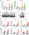
Fig. 1.
Fig. 1.
IRF5 forms a dimer and mediates cytokine induction by diverse pathways. (A) Depletion of IRF5 abolishes LPS-induced cytokine production in THP-1 cells. Cells used in this experiment included WT (THP-1 WT), IRF5 knockdown (THP-1 shIRF5), IRF5 knockdown rescued with mouse IRF5 (THP-1 shIRF5+Flag-mIRF5-HA, labeled THP-1 shIRF5+mIRF5), and IRF5 knockdown rescued with human IRF5 (THP-1 shIRF5+HA-hIRF5, labeled THP-1 shIRF5+hIRF5). These cells were stimulated with 5 µg/mL LPS for the indicated time before total RNA was isolated. CXCL10 and IL-12 (p40 subunit) mRNA levels were analyzed by qRT-PCR. Unless indicated otherwise, error bars represent SDs of triplicate assays. (B) IRF5 forms dimer on activation. The THP-1 shIRF5+Flag-mIRF5-HA cell line as described in A was left untreated (control; Ctl) or stimulated by incubation with LPS (5 µg/mL) or transfection with poly(dA:dT) (2 µg/mL), HT-DNA (2 µg/mL), or poly(I:C) (2 µg/mL) for the indicated time. The formation of IRF5 dimer was analyzed by native PAGE, followed by immunoblotting with the HA antibody. (C) IRF5 promotes cytokine induction in 293T cells. WT 293T cells and cells stably expressing Flag-mIRF5-HA were stimulated with Sendai virus (SeV) or poly(I:C) (2 µg/mL) for the indicated time, followed by measurement of TNF-α and IFN-β RNA levels by qRT-PCR. (Right) The cells were transfected with empty pcDNA vector, pcDNA-Flag-MAVS (MAVS), or pcDNA-Flag-IKKβ (IKKβ) for 24 h before total RNA was isolated for analysis by qRT-PCR. (D) Overexpression of IKKβ or MAVS activates IRF5 in cells. The 293T Flag-mIRF5-HA cell line as described in C was transiently transfected with empty pcDNA vector or the vector containing Flag-MAVS or Flag-IKKβ for 24 h. Dimerization of IRF5 was analyzed by native PAGE, followed by immunoblotting with the HA antibody.
Fig. 2.
Fig. 2.
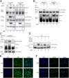
IKKβ activates IRF5 in vitro and in cells. (A and B) IKKβ activates IRF5 in vitro. (A) A cytosolic fraction (S20) from the 293T Flag-mIRF5-HA cell line was incubated with purified IKKβ or TBK1 protein in the presence of ATP. Dimerization of IRF5 or IRF3 was analyzed by native PAGE, followed by immunoblot analysis. (B) In vitro translated 35S-IRF5 or 35S-IRF3 protein was incubated with BSA, IKKβ, or TBK1 in the presence of ATP. Dimerization of IRF5 or IRF3 was analyzed by native PAGE, followed by autoradiography. Ctl, control cytosolic fraction without kinase. (C) IKKβ inhibitor blocks IRF5 activation by LPS. THP-1 shIRF5 cells stably reconstituted with Flag-mIRF5-HA were treated with IKKβ inhibitor (TPCA-1; 20 µM) or TBK1 inhibitor (BX-795; 10 µM) for 2 h before stimulation with LPS (5 µg/mL) for 2 h. IRF5 activation was analyzed by native PAGE and immunoblotting. Ctl, DMSO control. (D) Knockdown of IKKβ or TRAF6 abolishes IRF5 activation by MAVS. IKKα, IKKβ, TRAF6, or NEMO was stably knocked down in 293T Flag-mIRF5-HA cells using lentiviral shRNA as indicated. These cells were transfected with empty pcDNA vector, pcDNA-Flag-MAVS, or pcDNA-Flag-IKKβ for 24 h. (Upper) Activation of IRF5 was analyzed by native PAGE and immunoblot analysis. (Lower) Knockdown efficiency for each gene was analyzed by immunoblot analysis.
Fig. 3.
Fig. 3.
Mapping and functional analysis of IRF5 phosphorylation sites. (A) IKKβ activates IRF5 in vitro. IRF5 partially purified from 293T Flag-mIRF5-HA cells was incubated with IKKβ or BSA in the presence of ATP. Activation of IRF5 was analyzed by native PAGE and immunoblot analysis. (B) IKKβ phosphorylates IRF5 at Ser-445 and Ser-434. IRF5 in reaction mixtures described in A was purified with a Flag antibody and then analyzed by tandem mass spectrometry. The sequences of the peptides and number of nonphosphorylated and phosphorylated peptides in each condition are shown. (C) Representative tandem mass spectrum (MS2) after HCD fragmentation of the ion with m/z = 1061.50 (z = 2+) indicating phosphorylation at S445. “b” and “y” ions with or without neutral loss are labeled in blue. Diagnostic ions for phosphorylation are highlighted in red. (D) Serine 445 is essential for IRF5 activation by IKKβ in vitro. WT or mutant 35S-IRF5 proteins were translated in vitro and incubated with IKKβ or BSA in the presence of ATP. Dimerization of IRF5 was analyzed by native PAGE, followed by autoradiography. (E–G) Serine 445 of IRF5 is required for cytokine induction in cells. (E and F) 293T cell lines stably expressing WT or S445A IRF5 were transfected with expression vectors for IKKβ or MAVS for 24 h, or infected with Sendai virus for the indicated time. Total RNA was isolated for the measurement of TNF-α and IFNβ RNA levels by qRT-PCR. (G) WT (THP-1 WT), IRF5 knockdown (THP-1 shIRF5), and IRF5 knockdown and rescued with WT or S445A mouse IRF5 (THP-1 shIRF5+mIRF5 WT or THP-1 shIRF5+mIRF5 S445A) THP-1 cell lines were stimulated with 5 µg/mL LPS for 6 h before total RNA was isolated. IL-12 p40 mRNA levels were analyzed by qRT-PCR. *P < 0.05, a statistically significant difference.
Fig. 4.
Fig. 4.
IKKβ-dependent phosphorylation of IRF5 at Serine 445 in response to virus infection and LPS stimulation. (A) 293T cells stably expressing WT or S445A Flag-mIRF5-HA were transfected with expression vectors for IKKβ or MAVS for 24 h. (Upper) Aliquots of the cell extracts were analyzed for IRF5 dimerization by native PAGE, whereas other aliquots were immunoprecipitated with the HA antibody, followed by immunoblotting with an antibody against IRF5 or phosphorylated IRF5 at Ser-445. Expression of IKKβ and MAVS was examined by immunoblotting with the Flag antibody (Lower). (B) 293T cell lines as described above were treated with or without 20 µM TPCA-1 for 2 h before being infected with Sendai virus for 24 h. (Upper) IRF5 was immunoprecipitated with an HA antibody, followed by immunoblotting with an antibody against IRF5 or phosphorylated IRF5. Dimerization of IRF3 was detected by native PAGE and immunoblot analysis (Lower). (C) Raw 264.7 cell stably expressing Flag-mIRF5-HA was treated with or without TPCA-1 (20 µM) for 2 h before being stimulated with LPS (5 µg/mL) for 2 h. IRF5 was immunoprecipitated with an HA antibody, followed by immunoblotting with an antibody against IRF5 or phosphorylated IRF5. Dimerization of IRF5 was detected by immunoblotting of cytosolic extracts. (D) THP-1 cells were treated with or without the IKKβ inhibitors (TPCA-1 and PS1145) or TBK1 inhibitor (BX-795) for 2 h before stimulation with LPS (5 µg/mL) for 2 h. Phosphorylated IRF5 was immunoprecipitated with an IRF5 antibody, followed by immunoblotting with the same antibody or the phospho-IRF5 (S445) antibody. (E and F) Phosphorylated IRF5 accumulates in the nucleus. Differentiated THP-1 cells were stimulated with LPS for 2 h. Nuclear translocation and phosphorylation of IRF5 were monitored by confocal immunofluorescence using antibodies against IRF5 (E) or p-IRF5 (F).

Comment in

  • Innate sense of purpose for IKKβ.
    Hayden MS, Ghosh S. Hayden MS, et al. Proc Natl Acad Sci U S A. 2014 Dec 9;111(49):17348-9. doi: 10.1073/pnas.1419689111. Epub 2014 Nov 26. Proc Natl Acad Sci U S A. 2014. PMID: 25427798 Free PMC article. No abstract available.

Similar articles

Cited by

References

    1. Honda K, Taniguchi T. IRFs: Master regulators of signalling by Toll-like receptors and cytosolic pattern-recognition receptors. Nat Rev Immunol. 2006;6(9):644–658. - PubMed
    1. Tamura T, Yanai H, Savitsky D, Taniguchi T. The IRF family transcription factors in immunity and oncogenesis. Annu Rev Immunol. 2008;26:535–584. - PubMed
    1. Ikushima H, Negishi H, Taniguchi T. The IRF family transcription factors at the interface of innate and adaptive immune responses. Cold Spring Harb Symp Quant Biol. 2013;78:105–116. - PubMed
    1. Kawai T, et al. IPS-1, an adaptor triggering RIG-I- and Mda5-mediated type I interferon induction. Nat Immunol. 2005;6(10):981–988. - PubMed
    1. Meylan E, et al. Cardif is an adaptor protein in the RIG-I antiviral pathway and is targeted by hepatitis C virus. Nature. 2005;437(7062):1167–1172. - PubMed

Publication types