. 2014 Dec 1;592(23):5301-15.
doi: 10.1113/jphysiol.2014.280420. Epub 2014 Oct 17.

Ageing and muscular dystrophy differentially affect murine pharyngeal muscles in a region-dependent manner

Affiliations

Ageing and muscular dystrophy differentially affect murine pharyngeal muscles in a region-dependent manner

Matthew E Randolph et al. J Physiol. .

Abstract

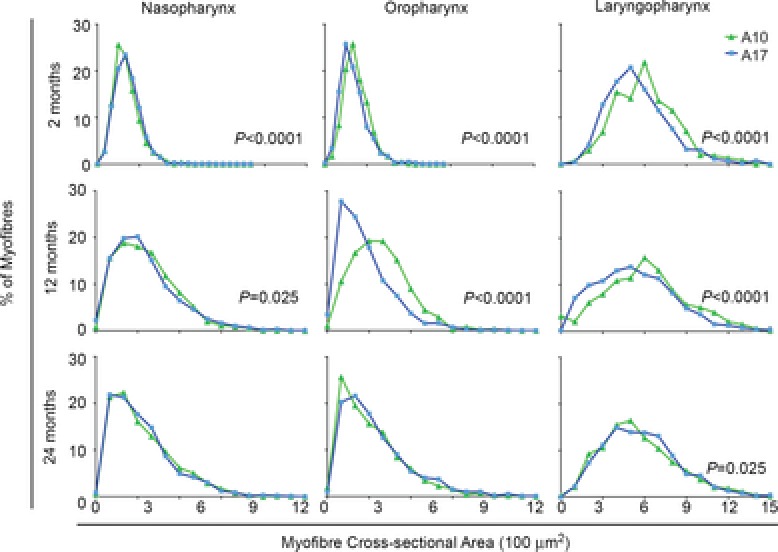
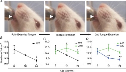
The inability to swallow, or dysphagia, is a debilitating and life-threatening condition that arises with ageing or disease. Dysphagia results from neurological or muscular impairment of one or more pharyngeal muscles, which function together to ensure proper swallowing and prevent the aspiration of food or liquid into the lungs. Little is known about the effects of age or disease on pharyngeal muscles as a group. Here we show ageing affected pharyngeal muscle growth and atrophy in wild-type mice depending on the particular muscle analysed. Furthermore, wild-type mice also developed dysphagia with ageing. Additionally, we studied pharyngeal muscles in a mouse model for oculopharyngeal muscular dystrophy, a dysphagic disease caused by a polyalanine expansion in the RNA binding protein, PABPN1. We examined pharyngeal muscles of mice overexpressing either wild-type A10 or mutant A17 PABPN1. Overexpression of mutant A17 PABPN1 differentially affected growth of the palatopharyngeus muscle dependent on its location within the pharynx. Interestingly, overexpression of wild-type A10 PABPN1 was protective against age-related muscle atrophy in the laryngopharynx and prevented the development of age-related dysphagia. These results demonstrate that pharyngeal muscles are differentially affected by both ageing and muscular dystrophy in a region-dependent manner. These studies lay important groundwork for understanding the molecular and cellular mechanisms that regulate pharyngeal muscle growth and atrophy, which may lead to novel therapies for individuals with dysphagia.


Figures

Figure 1
Figure 1. Pharyngeal muscles of mice
A, murine pharyngeal regions depicting the NP in yellow, OP in green, and LP in blue. B, representative histologic sections of murine pharyngeal tissue stained with haematoxylin and eosin. Pharyngeal muscles are outlined for identification. Representative images of the NP containing the superior pharyngeal constrictor (♦) and palatopharyngeus (*); the OP containing the middle pharyngeal constrictor (▴) and palatopharyngeus (*); and the LP containing the thyropharyngeus (•) and cricopharyngeus (▪) are shown. Bar: 250 μm. LP, laryngopharynx; NP, nasopharynx; OP, oropharynx.
Figure 2
Figure 2. Pharyngeal muscles are composed of fast glycolytic myofibres but lack slow oxidative myofibres
A, MHCs were isolated from the indicated muscles, separated by gel electrophoresis and visualized with coomassie blue (top panel). The main MHCs present in pharyngeal muscles (delineated within the black box) were types IIb and IIx, and neonatal based on comigration with MHC standards from other muscles. No type I MHC was observed in pharyngeal samples, which was confirmed by immunoblot analysis using the type I MHC-specific antibody, A4.84 (bottom panel). n = 4 mice pooled per sample. B, pharyngeal muscle sections were immunostained with antibodies against type I, IIa, IIx and IIb MHC. Representative sections from the palatopharyngeus and middle pharyngeal constrictor muscle are shown. Gastroc and soleus muscles were used as positive controls. Bar: 50 μm. n = 3 mice, 2–3 months of age. C, representative image of neonatal MHC immunostaining of the CP muscle in the LP marked with a dashed line to delineate the CP OL and CP IL of the muscle. Bar: 100 μm. n = 3 male mice, 2–3 months of age. CP IL, inner layers of the cricopharyngeal muscle; CP OL, outer layers of the cricopharyngeal muscle; EOM, extraocular muscle; F, female; Gastroc, gastrocnemius; LP, laryngopharynx; M, male; MHC, myosin heavy chain; mo, months; PE, pharyngeal epithelium; Phar, pharyngeal muscles.
Figure 3
Figure 3. Regional differences in myofibre size occur with ageing in pharyngeal muscles
Histogram plots are shown for myofibre cross-sectional areas (CSA) from the naso-, oro- and laryngopharyngeal regions of wild-type FVB mice at 2, 12 or 24 months of age. A, myofibre CSA significantly increased from 2 to 12 months of age in the naso- and oropharynx but decreased in the laryngopharynx. n = 752–1943 myofibres, three to four mice per time point. B, myofibre CSA decreased from 12 to 24 months of age in all pharyngeal regions. Data from 12 months of age are shown again for comparison. n = 792–1943 myofibres, four mice per time point. mo, months.
Figure 4
Figure 4. Overexpression of WT A10 PABPN1 enhances muscle growth in only one region of the pharynx
A, representative immunoblot of PABPN1 from pharyngeal muscle lysates of 2-month-old A10-WT, A17-MUT and WT littermates. Lanes were loaded with 100 μg total protein. Heat shock protein 90 was used as a loading control. Data are representative of three independent experiments. B, histogram plots are shown for myofibre CSA from the naso-, oro- and laryngopharyngeal regions of WT and A10-WT mice at 2, 12 or 24 months of age. Data from WT mice in Fig. 3 are shown again for comparison. At 2 months of age, overexpression of WT A10 PABPN1 significantly increased myofibre CSA in all three pharyngeal regions. At both 12 and 24 months of age, WT A10 PABPN1 overexpression only increased myofibre size in the laryngopharynx. P values are indicated in plots with significant differences in myofibre CSA. n = 609–1943 myofibres; three to five mice per genotype and time point. C, representative haematoxylin and eosin-stained section of the inferior constrictor muscle located in the laryngopharyx of a WT 2-month-old mouse. Centrally located myonuclei (black arrowheads) are present in multiple myofibres. Bar: 50 μm. D, quantification of centrally located myonuclei within the nasal, oral and laryngeal pharynxes of 2-month-old WT, A10-WT and A17-MUT mice. Centrally located myonuclei within A10-WT myofibres were significantly increased compared to WT (*P < 0.05) or A17-MUT (**P < 0.01) mice, but only in the laryngopharynx. Data are means ± SEM from 752 to 1214 myofibres from three mice per genotype. A10-WT, wild-type A10.1 PABPN1 overexpression transgenic mouse; A17-MUT, mutant A17.1 PABPN1 overexpression transgenic mouse; CSA, cross-sectional areas; LP, laryngeal pharynx; NP, nasal pharynx; OP, oral pharynx; PABN1, polyadenylate binding nuclear protein 1; WT, wild-type.
Figure 5
Figure 5. Overexpression of 17-alanine-expanded PABPN1 is deleterious to myofibre size only in specific regions of the pharynx
Histogram plots are shown for myofibre CSA from the naso-, oro- and laryngopharyngeal regions of A10-WT and A17-MUT mice at 2, 12 or 24 months of age. Data from A10-WT mice in Fig. 4 are shown again for comparison. At 2 months of age, myofibre CSA majorly decreased in the laryngopharynx of A17-MUT mice while CSA minimally changed in the naso- and oropharynx. At 12 months of age, major decreases in A17-MUT myofibre CSA occurred in the oro- and laryngopharynx. A pronounced lack of growth occurred in the A17-MUT oropharynx from 2 to 12 months. At 24 months of age, no major differences in myofibre CSA were observed in any pharyngeal region. P values are indicated in plots with significant differences in myofibre CSA. n = 609–2591 myofibres; three to five mice per genotype and time point. A10-WT, wild-type A10.1 PABPN1 overexpression transgenic mouse; A17-MUT, mutant A17.1 PABPN1 overexpression transgenic mouse; CSA, cross-sectional areas; PABN1, polyadenylate binding nuclear protein 1.
Figure 6
Figure 6. Overexpression of WT A10 PABPN1 protects against age- and muscular dystrophy-related dysphagia
A, single lick episode is depicted using still frames from a representative lick assay video. White arrowheads highlight the extension and retraction of the tongue. BD, quantification of lick rates of WT, A10-WT or A17-MUT mice at 6, 18 and 24 months of age. Data are means ± SEM from 3 to 13 mice. B, WT lick rates significantly decreased at 24 months of age (*P < 0.05). C, overexpression of WT A10 PABPN1 provided a protective effect on lick rates at both 18 and 24 months of age (#P < 0.05) when compared to WT mice at these time points. Data from WT and A10-WT mice. D, lick rates of A17-MUT mice significantly decreased at 18 and 24 months of age (*P < 0.05) and were significantly impaired at these time points compared to A10-WT mice (#P < 0.05). A10-WT, wild-type A10.1 PABPN1 overexpression transgenic mouse; A17-MUT, mutant A17.1 PABPN1 overexpression transgenic mouse; PABN1, polyadenylate binding nuclear protein 1; WT, wild-type.

References

    1. Aherne W, Ayyar DR, Clarke PA. Walton JN. Muscle fiber size in normal infants, children and adolescents. An autopsy study. J Neurol Sci. 1971;14:171–182. - PubMed
    1. Aloysius A, Born P, Kinali M, Davis T, Pane M. Mercuri E. Swallowing difficulties in Duchenne muscular dystrophy: indications for feeding assessment and outcome of videofluroscopic swallow studies. Eur J Paediatr Neurol. 2008;12:239–245. - PubMed
    1. Anvar SY, Raz Y, Verway N, van der Sluijs B, Venema A, Goeman JJ, Vissing J, van der Maarel SM, t Hoen PAC, van Engelen BGM. Raz V. A decline in PABPN1 induces progressive muscle weakness in oculopharyngeal muscle dystrophy and in muscle aging. Aging (Albany NY) 2013;5:412–426. - PMC - PubMed
    1. Apponi LH, Corbett AH. Pavlath GK. Control of mRNA stability contributes to low levels of nuclear poly(A) binding protein 1 (PABPN1) in skeletal muscle. Skelet Muscle. 2013;3:23. - PMC - PubMed
    1. Apponi LH, Leung SW, Williams KR, Valentini SR, Corbett AH. Pavlath GK. Loss of nuclear poly(A)-binding protein 1 causes defects in myogenesis and mRNA biogenesis. Hum Mol Genet. 2010;19:1058–1065. - PMC - PubMed

Publication types

MeSH terms

LinkOut - more resources