Review
. 2015 Feb;72(4):645-58.
doi: 10.1007/s00018-014-1763-4. Epub 2014 Oct 19.

Nectin family of cell-adhesion molecules: structural and molecular aspects of function and specificity

Affiliations
Review

Nectin family of cell-adhesion molecules: structural and molecular aspects of function and specificity

Dibyendu Samanta et al. Cell Mol Life Sci. 2015 Feb.

Abstract

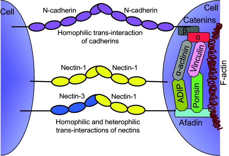
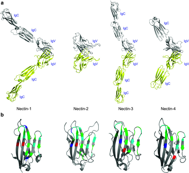
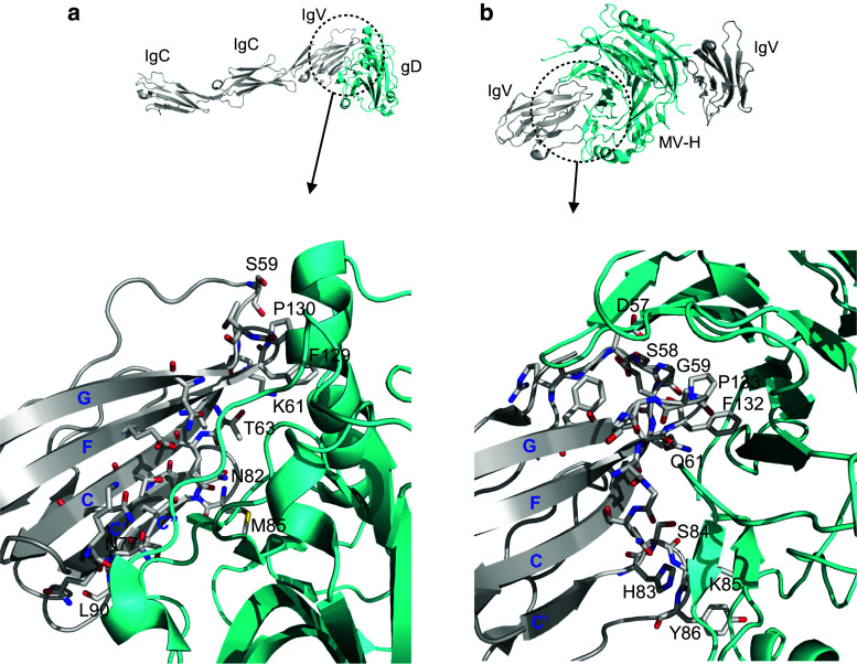
Cell-cell adhesive processes are central to the physiology of multicellular organisms. A number of cell surface molecules contribute to cell-cell adhesion, and the dysfunction of adhesive processes underlies numerous developmental defects and inherited diseases. The nectins, a family of four immunoglobulin superfamily members (nectin-1 to -4), interact through their extracellular domains to support cell-cell adhesion. While both homophilic and heterophilic interactions among the nectins are implicated in cell-cell adhesion, cell-based and biochemical studies suggest heterophilic interactions are stronger than homophilic interactions and control a range of physiological processes. In addition to interactions within the nectin family, heterophilic associations with nectin-like molecules, immune receptors, and viral glycoproteins support a wide range of biological functions, including immune modulation, cancer progression, host-pathogen interactions and immune evasion. We review current structural and molecular knowledge of nectin recognition processes, with a focus on the biochemical and biophysical determinants of affinity and selectivity that drive distinct nectin associations. These proteins and interactions are discussed as potential targets for immunotherapy.


Figures

Fig. 1
Fig. 1
Homophilic and heterophilic interactions of nectins. a Homophilic (black arrow) and heterophilic (red arrow) interactions among nectins are shown at the center. The homophilic affinities (equilibrium dissociation constant K d) of all the four nectins are stated. Nectins also participate in heterophilic interactions with a number of other proteins (shown in the periphery), including nectin-like molecules and some other immune receptors. b Recognition of nectin-2 by nectins, immune receptors (CD226, TIGIT) and viral glycoprotein D are important for cell–cell adhesion, immune modulation, and host-pathogen interactions, respectively
Fig. 2
Fig. 2
Domain organizations of the nectins and extended family members. Each nectin consists of three Ig-like domains (one IgV and two IgC) in their extracellular region, a single transmembrane (TM) region, and a cytoplasmic tail. The C-terminus of the cytoplasmic tail contains an afadin-binding motif
Fig. 3
Fig. 3
The interplay of nectins and cadherins at the site of cell–cell adhesions. Both nectin–afadin and cadherin-catenin systems work together and interact with F-actin bundles
Fig. 4
Fig. 4
Structural features of the nectins. a Structure of the IgV domain of human nectin-2 (a representative member of the family) showing the classical two-layer β-sandwich topology. The front and back sheets of the domain are composed of the GFCC′C″ and ABED strands, respectively. b Two monomers (yellow and gray) interact in a nearly orthogonal fashion to form the dimer interface, involving the front β sheets with predominant contribution from the C, C′, C″ and F strands of each monomer. c Key residues of the dimer interface of nectin-2 are represented by ball-and-stick representation. d Structure-based sequence alignment of the human nectins. The secondary structure of the nectin-2 IgV domain is displayed on the top of the alignment. Residues with similar properties are marked by red color, whereas identical residues are colored in white with red background. Interfacial residues (green circles) and the cysteine residues involved in the formation of disulfide bond between B and F strand (blue circle) are marked. The long loop between the D and E strand of nectin-2, which is absent in other nectins, is also shown
Fig. 5
Fig. 5
Ribbon representations of human nectin homodimers showing the overall structural organizations and interfacial residues at the dimer interfaces. a Individual protomers are colored in yellow and grey. The presented structures of nectin-1 (3ALP) and nectin-3 (4FOM) contain all the three extracellular domains, while the structure of nectin-2 (3R0N) has only the IgV and the structure of nectin-4 (4FRW) has the IgV and only a single IgC. b Ribbon diagram of the IgV domains representing the interfacial residues involved in the homodimerization of each nectin. The residues in red/blue represent charged residues, green represents polar residues, and cyan represents hydrophobic residues
Fig. 6
Fig. 6
Ribbon representations of homophilic and heterophilic interfaces of nectins. a Homodimer interface of human nectin-2 (PDB ID: 3R0N) showing the close proximity of two negatively charged side chains; Glu-141 is contributed from the F strand of each monomer (yellow and gray). b Dimeric interface of nectin-1 (PDB ID: 3ALP) showing the similar unfavorable repulsive electrostatics as depicted in case of nectin-2. c Molecular model showing the heterodimer interface of nectin-2 (yellow) and nectin-3 (brown). The modeling suggests that E141 of nectin-2 contacts K149 of nectin-3, forming a putative polar interaction at the center of the dimer interface which favors a strong heterophilic interaction
Fig. 7
Fig. 7
Schematic comparison of CD226/TIGIT:nectin-2/PVR and CD28/CTLA-4:B7-1/B7-2 networks. CD226 is a putative activating receptor-like CD28, whereas TIGIT is an inhibitory receptor-like CTLA-4. Both sets of molecules are engaged by sets of counter-ligands, TIGIT and CD226 by nectin-2 and PVR, and CTLA-4 and CD28 by B7-1 and B7-2. Activation is indicated by ‘+’ and inhibition is indicated by ‘–’ symbol
Fig. 8
Fig. 8
Crystal structure of nectin-1 in complex with HSV-1 gD (3U82) and nectin-4 with MV-H (4GJT). a A ribbon representation of nectin-1/gD complex structure (nectin-1 in grey and gD in cyan) showing the interacting residues of nectin-1 that are distributed through out the front sheet (C, C′, C″, F, and G strands); most of them are identical to the residues required for nectin-1 homodimerization. b Ribbon representation of nectin-4 IgV/MV-H complex structure (nectin-4 in grey and MV-H in cyan) showing the interacting residues of nectin-4 that are localized in C′–C″, B–C and F–G loops, which are distinct from the residues involved in the homodimerization of nectin-4

References

    1. Yap AS, Brieher WM, Gumbiner BM. Molecular and functional analysis of cadherin-based adherens junctions. Annu Rev Cell Dev Biol. 1997;13:119–146. doi: 10.1146/annurev.cellbio.13.1.119. - DOI - PubMed
    1. Takai Y, Ikeda W, Ogita H, Rikitake Y. The immunoglobulin-like cell adhesion molecule nectin and its associated protein afadin. Annu Rev Cell Dev Biol. 2008;24:309–342. doi: 10.1146/annurev.cellbio.24.110707.175339. - DOI - PubMed
    1. Takai Y, Miyoshi J, Ikeda W, Ogita H. Nectins and nectin-like molecules: roles in contact inhibition of cell movement and proliferation. Nat Rev Mol Cell Biol. 2008;9(8):603–615. doi: 10.1038/nrm2457. - DOI - PubMed
    1. Harris TJ, Tepass U. Adherens junctions: from molecules to morphogenesis. Nat Rev Mol Cell Biol. 2010;11(7):502–514. doi: 10.1038/nrm2927. - DOI - PubMed
    1. Sakisaka T, Takai Y. Biology and pathology of nectins and nectin-like molecules. Curr Opin Cell Biol. 2004;16(5):513–521. doi: 10.1016/j.ceb.2004.07.007. - DOI - PubMed

Publication types

MeSH terms