. 2015 Jan;33(1):102-6.
doi: 10.1038/nbt.3055. Epub 2014 Oct 19.

In vivo interrogation of gene function in the mammalian brain using CRISPR-Cas9

Affiliations

In vivo interrogation of gene function in the mammalian brain using CRISPR-Cas9

Lukasz Swiech et al. Nat Biotechnol. 2015 Jan.

Abstract

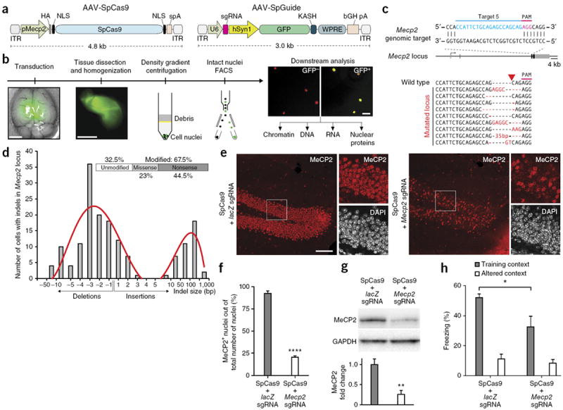
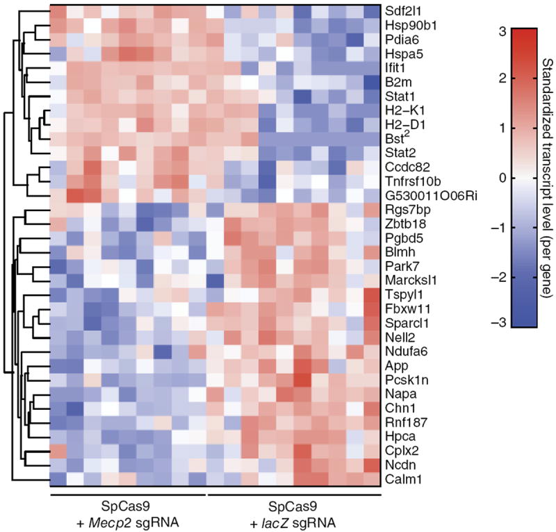
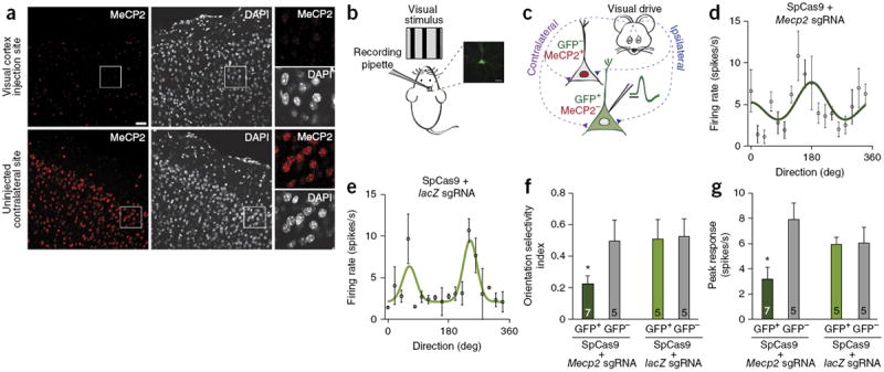
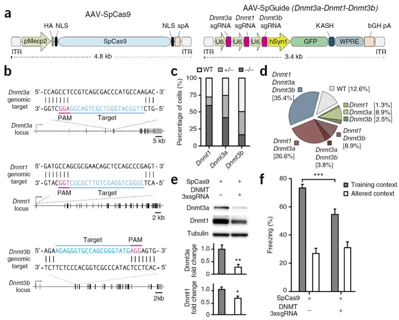
Probing gene function in the mammalian brain can be greatly assisted with methods to manipulate the genome of neurons in vivo. The clustered, regularly interspaced, short palindromic repeats (CRISPR)-associated endonuclease (Cas)9 from Streptococcus pyogenes (SpCas9) can be used to edit single or multiple genes in replicating eukaryotic cells, resulting in frame-shifting insertion/deletion (indel) mutations and subsequent protein depletion. Here, we delivered SpCas9 and guide RNAs using adeno-associated viral (AAV) vectors to target single (Mecp2) as well as multiple genes (Dnmt1, Dnmt3a and Dnmt3b) in the adult mouse brain in vivo. We characterized the effects of genome modifications in postmitotic neurons using biochemical, genetic, electrophysiological and behavioral readouts. Our results demonstrate that AAV-mediated SpCas9 genome editing can enable reverse genetic studies of gene function in the brain.


Conflict of interest statement

COMPETING FINANCIAL INTERESTS

The authors declare competing financial interests: details are available in the online version of the paper.

Figures

Figure 1
Figure 1
Targeting of Mecp2 locus in the adult mouse brain with SpCas9. (a) AAV-SpCas9 and AAV-SpGuide expression vectors. The sgRNA vector contains encoding sequence of the GFP-KASH fusion protein for identification of transduced neurons. (b) Strategy for cell nuclei purification of CRISPR-Cas9 targeted cells from the mouse brain. Scale bars: 3 mm (brain), 50 μm (sorted nuclei). (c) Graphical representation of the mouse Mecp2 locus showing SpCas9 target location; targeted genomic locus indicated in blue. PAM sequence marked in magenta. Representative mutation patterns detected by sequencing of Mecp2 locus shown below: top, wild-type sequence; red dashes, deleted bases; red bases: insertion or mutations (indel); red arrowhead indicates CRISPR-Cas9 cutting site. (d) Indel frequency in SpCas9 targeted Mecp2 locus (240 sorted nuclei from dissected hippocampi; n = 4 male mice, 2 weeks after AAV injection). Fraction of missense and nonsense mutations is shown. Distribution of indel length in single sorted nuclei is shown on the bar graph below. (e) Immunostaining of dorsal DG region 2 weeks after CRISPR-Cas9 targeting of Mecp2 locus in male mice. Scale bar, 150 μm. (f) Quantification of MeCP2 positive cells population within all detected cells (DAPI staining) in DG compare to control (t-test, ****P < 0.0001, n = 290 and 249 cells from 2 male mice, respectively; error bars: s.e.m.). (g) Western blot analysis of MeCP2 protein expression 2 weeks after AAV injection and quantification of MeCP2 protein levels in dorsal DG (t-test, **P < 0.01, n = 4 tissue punches from male mice, error bars: s.e.m.). (h) Contextual learning deficits after targeting Mecp2 using SpCas9 in the dorsal DG region of hippocampus, tested in training and altered context (t-test, *P < 0.05, n = 7 male mice, 3 weeks after AAV delivery; error bars: s.e.m.). ITR, inverted terminal repeat; HA, hemagglutinin tag; NLS, nuclear localization signal; spA, synthetic polyadenylation signal; U6, Pol III promoter; sgRNA, single guide RNA; hSyn, human synapsin 1 promoter; GFP, green fluorescent protein; KASH, Klarsicht, ANC1, Syne Homology nuclear transmembrane domain; bGH pA, bovine growth hormone polyadenylation signal; WPRE, Woodchuck Hepatitis virus posttranscriptional regulatory element.
Figure 2
Figure 2
Analysis of gene expression in SpCas9-mediated MeCP2 knockdown neurons. Hierarchical clustering of differentially expressed genes (t-test, 0.01 FDR, n = 19 populations of sorted nuclei from 8 male mice, 2 weeks after AAV delivery) detected by RNA-seq. Relative log(TPM+1) expression levels of genes are normalized for each row and displayed in red-blue color scale. Each column represents a population of targeted 100 neuronal nuclei FACS sorted from the isolated, dentate gyrus population of cells, either from Mecp2 or control (lacZ) sgRNA transduced animals, as indicated. TPM, transcripts per million.
Figure 3
Figure 3
Changes in response properties of visual cortex neurons after SpCas9-mediated MeCP2 knockdown. (a) Immunostaining of cortex (V1), 2 weeks after CRISPR-Cas9 targeting of Mecp2 locus in male mice. Virus injected site, and uninjected control contralateral side are shown. Scale bar, 50 μm. (b) Experimental configuration. To assess visual responses of neurons in primary visual cortex (V1), we presented visual stimuli (oriented gratings) on an LCD monitor placed in front of anesthetized mice. Two weeks before experiment, mixture of SpCas9 with either Mecp2 sgRNA or control sgRNA (lacZ) was stereotactically injected in V1. GFP-KASH+ neurons and GFP-KASH neurons were recorded. Example of recorded GFP-KASH+ neuron is shown. Scale bar, 20 μm. (c) In vivo targeted cell-attached recording configuration from V1 layer 2/3 excitatory neurons that receive visual drive (ipsilateral and contralateral input). Genome modified GFP-KASH+/MeCP2 cells are shown in green, unmodified GFP-KASH/MeCP2+ cells in gray. Recording pipette is indicated. Normalized mean spike shape (green) shows regular spiking excitatory neurons. Scale bar, 1 ms. (d, e) Orientation-selective responses of typical GFP-KASH+ neurons expressing Mecp2 sgRNA (d) or lacZ sgRNA (e). (f, g) Orientation selectivity index (f) and peak responses (g) (spikes/s) measured from GFP-KASH+ cells expressing Mecp2 and control sgRNA, respectively, and comparison with GFP-KASH cells (*P < 0.05, t-test; numbers on bars indicate numbers of recorded cells; n = 2–3 animals per group; error bars: s.e.m.).
Figure 4
Figure 4
Simultaneous, multiplex gene editing in the mouse brain. (a) Schematic illustration of AAV vectors for multiplex genome targeting. (b) Graphical representation of targeted DNMT mouse loci. Targeted genomic loci are indicated in blue. PAM sequences are marked in magenta. (c) Next-generation sequencing-based analysis of indel frequency formation within Dnmt1, Dnmt3a and Dnmt3b loci analyzed in single neuronal nuclei. Fractions of mono- and biallelic modification are shown (n = 79 cells). (d) NGS analysis of DNMTs loci modification in single cells, showing co-occurrence of modification in multiple loci. (e) Western blot analysis for Dnmt3a and Dnmt1 proteins after in vivo delivery of CRISPR-Cas9 system targeting DNMT family genes in neurons (top). Western blot quantification of Dnmt3a and Dnmt1 protein levels in DG after in vivo CRISPR-Cas9 targeting (bottom; t-test, **P < 0.001, *P < 0.05, Dnmt3a: n = 7; Dnmt1: n = 5 from 5 animals; error bars: s.e.m.). (f) Contextual learning deficits, 8 weeks after targeting of DNMT genes using SpCas9 in the DG region of hippocampus, tested in training and altered context (t-test, ***P < 0.001, n = 18 animals; error bars: s.e.m.).

Similar articles

Cited by

References

    1. Hsu PD, Lander ES, Zhang F. Development and applications of CRISPR-Cas9 for genome engineering. Cell. 2014;157:1262–1278. - PMC - PubMed
    1. Xue W, et al. CRISPR-mediated direct mutation of cancer genes in the mouse liver. Nature. 2014 Aug 6; doi: 10.1038/nature13589. - DOI - PMC - PubMed
    1. Yin H, et al. Genome editing with Cas9 in adult mice corrects a disease mutation and phenotype. Nat Biotechnol. 2014;32:551–553. - PMC - PubMed
    1. Ding Q, et al. Permanent alteration of PCSK9 with in vivo CRISPR-Cas9 genome editing. Circ Res. 2014;115:488–492. - PMC - PubMed
    1. Wu Z, Yang H, Colosi P. Effect of genome size on AAV vector packaging. Mol Ther. 2010;18:80–86. - PMC - PubMed

Publication types

MeSH terms

Associated data