. 2015 Jan 8;517(7533):223-6.
doi: 10.1038/nature13826. Epub 2014 Oct 19.

Allosteric activation of the RNF146 ubiquitin ligase by a poly(ADP-ribosyl)ation signal

Affiliations

Allosteric activation of the RNF146 ubiquitin ligase by a poly(ADP-ribosyl)ation signal

Paul A DaRosa et al. Nature. .

Abstract

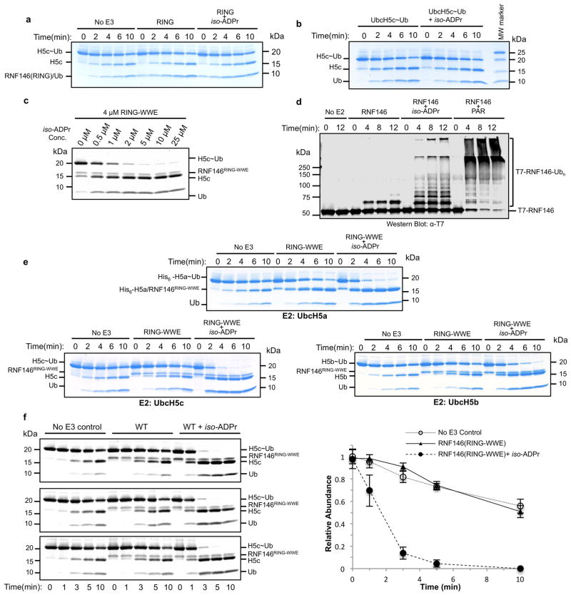
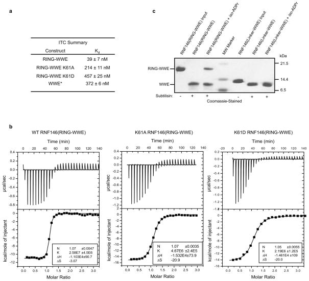
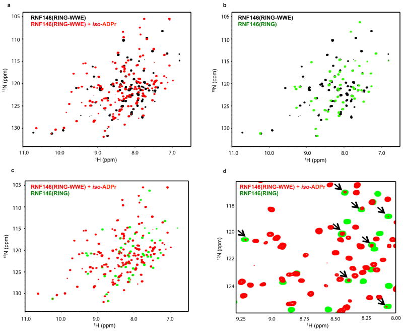
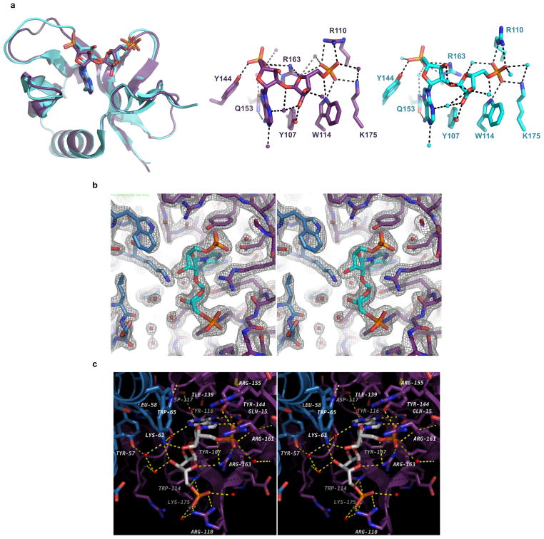
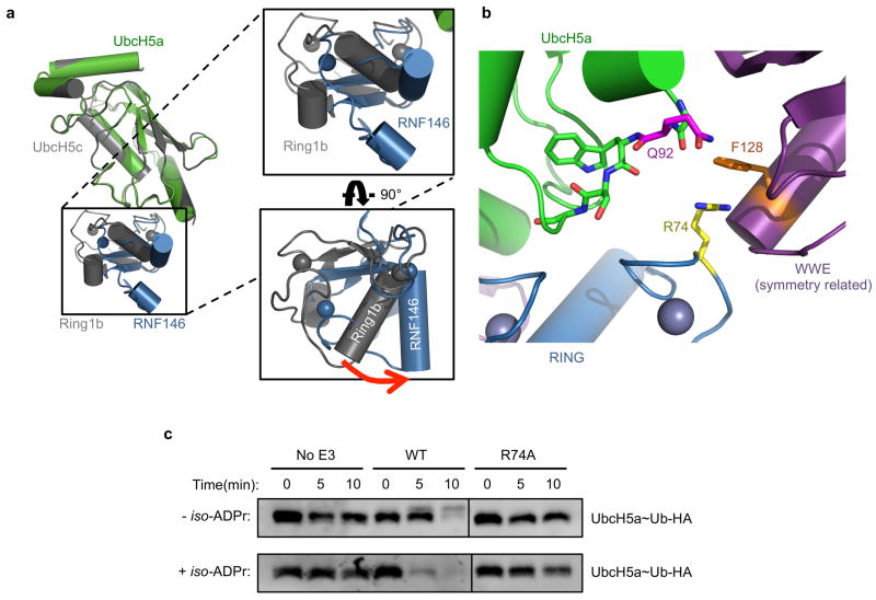
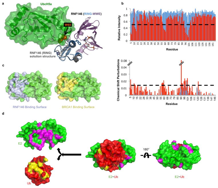
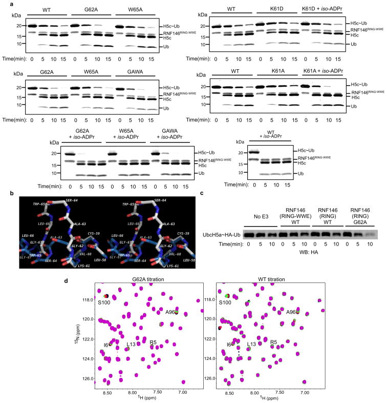
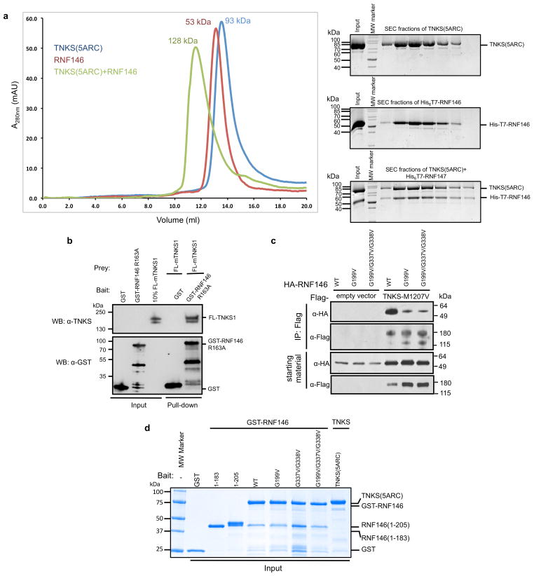
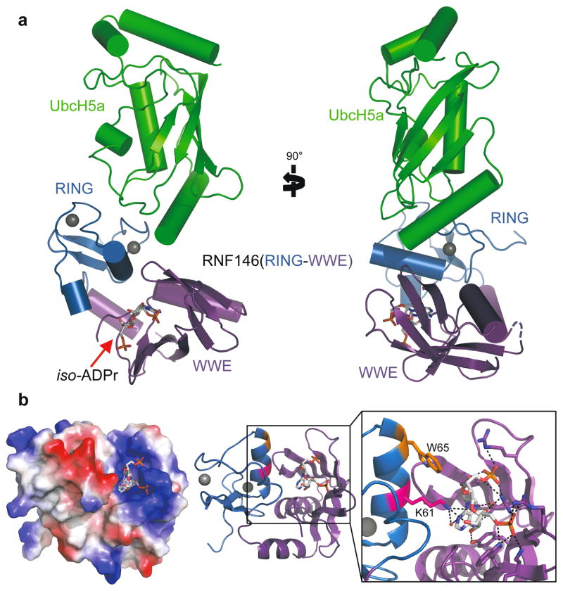
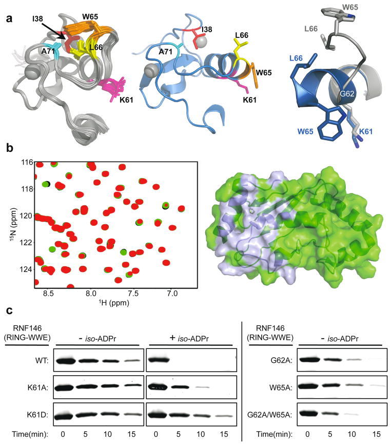
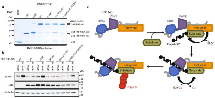
Protein poly(ADP-ribosyl)ation (PARylation) has a role in diverse cellular processes such as DNA repair, transcription, Wnt signalling, and cell death. Recent studies have shown that PARylation can serve as a signal for the polyubiquitination and degradation of several crucial regulatory proteins, including Axin and 3BP2 (refs 7, 8, 9). The RING-type E3 ubiquitin ligase RNF146 (also known as Iduna) is responsible for PARylation-dependent ubiquitination (PARdU). Here we provide a structural basis for RNF146-catalysed PARdU and how PARdU specificity is achieved. First, we show that iso-ADP-ribose (iso-ADPr), the smallest internal poly(ADP-ribose) (PAR) structural unit, binds between the WWE and RING domains of RNF146 and functions as an allosteric signal that switches the RING domain from a catalytically inactive state to an active one. In the absence of PAR, the RING domain is unable to bind and activate a ubiquitin-conjugating enzyme (E2) efficiently. Binding of PAR or iso-ADPr induces a major conformational change that creates a functional RING structure. Thus, RNF146 represents a new mechanistic class of RING E3 ligases, the activities of which are regulated by non-covalent ligand binding, and that may provide a template for designing inducible protein-degradation systems. Second, we find that RNF146 directly interacts with the PAR polymerase tankyrase (TNKS). Disruption of the RNF146-TNKS interaction inhibits turnover of the substrate Axin in cells. Thus, both substrate PARylation and PARdU are catalysed by enzymes within the same protein complex, and PARdU substrate specificity may be primarily determined by the substrate-TNKS interaction. We propose that the maintenance of unliganded RNF146 in an inactive state may serve to maintain the stability of the RNF146-TNKS complex, which in turn regulates the homeostasis of PARdU activity in the cell.


Conflict of interest statement

Competing financial interests

The authors declare no competing financial interests.

Figures

Extended Data Figure 1
Extended Data Figure 1. Multiple sequence alignment (MSA) of RNF146 orthologs
The colored bars above the sequence alignment indicate regions of interest in human RNF146: RING domain (blue), WWE domain (purple), and potential TNKS binding motifs, numbered I through V (orange). While there are no apparent RXXGDG TNKS binding motifs, the five potential binding motifs indicated here are based on the TNKS-substrate interface plasticity demonstrated by a recent crystal structure of the Axin-TNKS complex.
Extended Data Figure 2
Extended Data Figure 2. Both PAR and iso-ADPr can activate RNF146 E3 ligase activity
a, Coomassie-stained E2~Ub/lysine reactivity of the RNF146 RING domain with and without iso-ADPr. The RING domain does not enhance E2~Ub conjugate reactivity in the absence or presence of ligand. b, Intrinsic lysine reactivity of the UbcH5c~Ub conjugate with and without iso-ADPr. iso-ADPr does not enhance the reactivity of the conjugate in the absence of RNF146. c, Oriole-stained E2~Ub/lysine reactivity with increasing iso-ADPr (3 min after lysine addition). The rate of E2~Ub/lysine reactivity is increased as a function of [iso-ADPr] up to 5 μM ligand addition (1.2 equiv.), consistent with the affinity of RNF146 for iso-ADPr (see Extended Data Fig. 3). d, Auto-ubiquitination of full-length RNF146 in the absence or presence of iso-ADPr or PAR polymer. Image shows western blot for T7-tagged RNF146. Because full-length RNF146 and the RING-WWE fragment have similar abilities to enhance E2~Ub reactivity (see Figure 1), the additional auto-ubiquitination seen with PAR is likely due to increased local concentration of RNF146 near PAR polymers, allowing auto-ubiquitination in trans. e, E2/lysine reactivity of UbcH5a, UbcH5b, and UbcH5c ubiquitin conjugates with RNF146(RING-WWE) in the absence or presence of iso-ADPr (Coomassie-stained). All three isoforms function with ligand-activated RNF146. f, Technical triplicates of RNF146(RING-WWE) E2~Ub/lysine reactivity assays (Oriole-stained; left) and a plot of relative densitometry values of the E2~Ub conjugate (right). Error bars indicate the mean +/- one standard deviation from three separate experiments. All times are given in minutes. “No E3” samples do not contain RNF146.
Extended Data Figure 3
Extended Data Figure 3. Both RNF146 RING and WWE domains contribute to iso-ADPr binding
a, Summary of iso-ADPr binding (Kd values) for RNF146(RING-WWE) obtained from the ITC titrations in the current work, and for the WWE-only fragment (previously published; indicated by asterisk). These data indicate that the RING domain contributes to iso-ADPr binding. b, Raw ITC titrations of RNF146(RING-WWE) fragments: (left to right) WT, K61A, and K61D. c, Limited proteolysis of RNF146(RING-WWE) and of a construct of RNF146 including the linker between the RING and WWE domains, and the WWE domain (RNF146(Linker-WWE); residues 83–183). Both appear to result in the same product when treated with subtilisin. The RING-WWE construct is more resistant to subtilisin in the presence of ligand.
Extended Data Figure 4
Extended Data Figure 4. 1H-15N HSQC-TROSY spectra of RNF146 reveal a conformational change in the RING domain upon iso-ADPr binding
a, RNF146(RING-WWE) spectra in the absence (black) and presence (red) of saturating iso-ADPr concentrations show a dramatic change in a majority of amide chemical environments. b, Overlay of the RNF146(RING-WWE) spectrum (black) with the spectrum of the RNF146 RING-only domain (green) in the absence of iso-ADPr. Nearly all RING-only peaks overlay with a peak in the RING-WWE fragment spectrum, confirming that the RING-only domain adopts the same conformation as the RING domain in the larger fragment. c, Overlay of the liganded RING-WWE spectrum (red) with the isolated RING domain (green) shows very few corresponding peaks between the two spectra, indicating environment changes of most RING domain peaks in the presence of iso-ADPr consistent with a conformational change. Importantly, there are no changes in the spectrum of the RING-only construct when iso-ADPr is added under these conditions (data not shown). d, Close-up of panel c to illustrate that the RING domain samples a minor conformational state in liganded RNF146(RING-WWE). The minor peaks all correspond to RING peaks of unliganded RNF146 (black arrows). Therefore, the RING domain can still sample the non-activated conformation when saturated with ligand. Spectra were obtained with 200 μM protein and 300 μM iso-ADPr (saturating conditions).
Extended Data Figure 5
Extended Data Figure 5. Comparison of ligand binding in the RNF146(RING-WWE)/UbcH5a/iso-ADPr complex and in the WWE-only structure
a, (Top left) Superposition of the WWE domain of the RNF146(RING-WWE)/UbcH5a/iso-ADPr complex (purple) and the previous iso-ADPr/WWE structure (cyan, PDB 3V3L). (Top center) WWE residues involved in binding iso-ADPr in the RNF146(RING-WWE)/UbcH5a/iso-ADPr complex (purple) and (Top right) in the previous iso-ADPr/WWE structure (cyan). Waters are shown as non-bonded spheres; hydrogen bonds are shown as dashed lines. Side-chain contacts between ligand and protein are maintained in both structures. b, Stereoview of the RNF146(RING-WWE)/UbcH5a/iso-ADPr complex ligand binding site showing the 2Fo-Fc map (grey mesh) contoured at 1.5 s. The ligand and waters are well defined within the binding site. Waters are shown as red non-bonded spheres, iso-ADPr is shown in cyan, and the RING and WWE domains are colored as in Fig. 2a. c, Stereoview of the iso-ADPr binding site indicating residues within 4.5 Å of the ligand. Protein and ligand are represented as sticks, waters as red non-bonding spheres, and hydrogen bonds as dashed yellow lines. The RING and WWE domains and ligand are colored as in Fig. 2a.
Extended Data Figure 6
Extended Data Figure 6. Rotation and crystal packing at the E2-E3 binding interface of the RNF146(RING-WWE)/UbcH5a/iso-ADPr complex
a, Superposition of the E2 in the RNF146(RING-WWE)/UbcH5a/iso-ADPr (colored as in Figure 2a) with a representative RING E3-E2 structure, the Bmi1/Ring1b-UbcH5c complex (grey) (PDB 3RPG). The WWE domain is excluded for clarity. Boxes show close-up views of the RING domains revealing a rotation of the RING domain relative to the E2. (Bottom right) RING domains rotated 90° to show the E2 binding surface of the E3s. The RING of the RNF146(RING-WWE)/UbcH5a/iso-ADPr structure is rotated relative to Ring1b-UbcH5c and other E3-E2 complexes–, (indicated by red arrow) when the E2s are aligned. b, A close-up view of the E2-E3 interface of the RNF146(RING-WWE)/UbcH5a/iso-ADPr complex shows that RING residue R74 (yellow) is too far (~7.7 Å) from the E2 Q92 (magenta) carbonyl to make the hydrogen bond observed in activated E3-E2~Ub structures,,,. The side chain of R74 in the RING domain packs against F128 (orange), a WWE domain residue of a symmetry-related molecule in this crystal form. It is likely that crystal packing interferes with the formation of the “allosteric” hydrogen bond. c, E2~Ub/lysine reactivity with RNF146(RING-WWE)-R74A shows a dependence of RNF146 activity on the allosteric arginine,,, with or without ligand. Because RNF146 activation requires R74, which does not make contacts with the E2 in our structure, and because RNF146 shows canonical E2 binding in solution (see Extended Data Fig. 7), we conclude that the orientation of RNF146 in the RNF146(RING-WWE)/UbcH5a/iso-ADPr complex is likely an unproductive E2-E3 association. The observed rotation is likely a crystallographic artifact.
Extended Data Figure 7
Extended Data Figure 7. RNF146/iso-ADPr binding allows the RING domain to bind and activate a ubiquitin conjugating enzyme (E2)
a, (Left) Superposition of the RING domain of unliganded RNF146 (PDB 2D8T; grey, Trp 65 is shown as orange spheres) with the RNF146(RING-WWE)/UbcH5a/iso-ADPr complex (colored as in Fig 2a), shows a clash of Trp 65 with UbcH5a at the E2-E3 binding interface. This clash is observed when the RNF146 RING structure (2D8T) is aligned with all other E2-E3 structures–,. b, Peak broadening (Top; intensity relative to free E2) and chemical shift perturbations (CSPs; bottom) of 15N-UbcH5c(S22R/C85S) resonances (data are from the spectra shown in Figure 3b). Histograms shown in blue compare the spectral properties of free E2 to E2 plus RNF146(RING-WWE); histograms shown in red compare free E2 to E2 plus RNF146(RING-WWE) and iso-ADPr. Dashed lines indicate one standard deviation from the mean value of the liganded (red) plots. Values below and above the dashed lines for the relative intensities and CSPs respectively are plotted on the E2 surface shown in panel c and Figure 3b. c, (Left) The RNF146(RING-WWE) binding surface inferred from data in panel b (light blue, on green E2), is compared with (right) the BRCA1/BARD1 binding surface on E2 (yellow, on green E2; residues 1–112 and residues 26–115, respectively) previously inferred by an analogous experiment. When the NMR perturbations are mapped to the surface of UbcH5c, the revealed binding sites are very similar, and are consistent with previously reported binding surfaces for RING E3s on free ubiquitin conjugating enzymes–,. d, Chemical shift perturbations and broadening of resonances from 15N-E2~Ub conjugate (UbcH5c(S22R/C85S)-O-Ub) upon RNF146(RING-WWE)/iso-ADPr binding (determined by the same method as shown in panel b, but with only 0.125 mol. equiv. E3 added to minimize hydrolysis of the E2~Ub oxyester during NMR data collection). (Left) Perturbed residues are mapped onto UbcH5b (magenta on green E2) and ubiquitin (yellow on red ubiquitin). (Center and right) Perturbed residues mapped onto the structure of E2~Ub as it appears in the E3/E2~Ub complex of BIRC7/UbcH5b-Ub (PDB 4AUQ; BIRC7 not shown for clarity) show that the surfaces highlighted in the left panel are buried in the “closed” state. The data show that RNF146 activates the E2~Ub conjugate by inducing the closed conformation,,,. Because only the most perturbed residues are mapped to the E2~Ub surface, the E3 binding surface is not highlighted on the E2 in panel d.
Extended Data Figure 8
Extended Data Figure 8. Stabilizing Helix 1 of RNF146 activates the RING domain
a, Complete images of gels shown in Figure 3c (Oriole-stained) for G62A, W65A, G62A/W65A (GAWA), K61A and K61D mutants of RNF146(RING-WWE) with and without iso-ADPr. G62A and GAWA mutants show reduced enhancement with iso-ADPr relative to wild type, likely due to a clash of the Ala side chain with a turn in the WWE domain at position 62 (data not shown). b, Alignment of RNF146(RING) solution structure (PDB 2D8T; white) and the crystal structure determined in this study (blue) shown in stereoview. Side chains are excluded for clarity; the backbone is represented by sticks. Comparison of the conformation of Gly 62 in the two structures suggests a need for a small side chain at position 62 to allow the structural transition from the inactive to active form of RNF146. c, Anti-HA western blot of the E2~Ub/lysine reactivity assay of RNF146(RING-WWE) compared with RNF146(RING) and RNF146(RING)-G62A showing enhanced reactivity for the G to A mutation. d, (Left) 1H-15N HSQC TROSY of 15N-UbcH5c(S22R/C85S) in the presence of 0.0 (black), 0.25 (red), 0.5 (green), and 1.0 (magenta) mol. equiv. of RNF146(RING) G62A. (Right) The same experiment performed with WT RNF146(RING). The most perturbed residues, indicated by letter and position (S100, etc.), show increased chemical shift perturbations for the RNF146(RING)-G62A mutant.
Extended Data Figure 9
Extended Data Figure 9. RNF146 directly interacts with TNKS
a, (Left) SEC profiles of untagged TNKS(5ARC) (blue), His6T7-RNF146 (red), and a 1:1 mixture of these proteins (green). Numbers above the peaks indicate the average mass obtained by multi-angle static light scattering (MALS) for each peak. His6T7-RNF146 and TNKS(5ARC) co-migrate as a single peak with an apparent mass of 128 kDa. (Right) Coomassie-stained SDS-PAGE analysis of the SEC peaks in left panel show the presence of both proteins within the peak of the TNKS(5ARC)/His6T7-RNF146 complex (Bottom right). b, GST pull-down of partially purified full-length mouse tankyrase-1 (FL-mTNKS1) with GST tagged RNF146-R163A (PAR-binding deficient RNF146 mutant). Full-length mTNKS1 can be pulled down by GST-RNF146, but not GST. c, Co-immunoprecipitation of HA-RNF146 variants with transiently transfected flag-tagged TNKS-M1207V (catalytically inactive mutant). The M1207V mutation prevents auto-PARylation of TNKS and therefore PAR-mediated interactions between RNF146 and TNKS. Under the experimental conditions, both the motif I mutant, G199V, and the motifs I+IV mutant, G199V/G337V/G338V, significantly reduce the RNF146-TNKS interaction. d, Coomassie-stained SDS-PAGE of proteins used in the GST pull-down assay shown in Figure 4a (inputs). Samples were used in a 1:2 ratio (3 μM GST-RNF146 to 6.7 μM TNKS(5ARC)) for these GST pull-down experiments.
Figure 1
Figure 1. Binding of iso-ADPr or PAR activates RNF146 E3 ubiquitin ligase activity
a. Domain structure of RNF146, with constructs used in this study shown: RNF146(RING), residues 30–89; RNF146(RING-WWE), residues 30–183. Five potential TNKS-binding motifs are shown in orange and numbered I through V in Roman numerals. b. Coomassie-stained E2~Ub/lysine reactivity assays of full-length RNF146 (top) and RNF146(RING-WWE) (bottom) with the E2 UbcH5c. Active RING domains enhance the reactivity of E2~Ub with lysine and therefore speed the disappearance of the E2~Ub species with the corresponding appearance of free E2 and Ub. The WWE domain does not bind ADP-ribose (ADPr).
Figure 2
Figure 2. Crystal structure of the RNF146(RING-WWE)/UbcH5a/iso-ADPr complex
a. Cartoon representation of the RNF146/UbcH5a complex with RING domain colored blue, WWE domain colored purple, and UbcH5a colored green. Zn2+ ions are shown as grey spheres, and the iso-ADPr ligand is represented as sticks. b. The RNF146/iso-ADPr interface. (Left) Surface electrostatic view of RNF146(RING-WWE), showing the iso-ADPr/PAR binding pocket; (Center) same view, cartoon representation; (Right) close-up view of iso-ADPr pocket. Polar contacts between protein and the ligand, iso-ADPr, are indicated by dashed lines; RING residues K61 (magenta) and W65 (orange) are highlighted.
Figure 3
Figure 3. Mechanism of RNF146 PAR-mediated RING activation
a, (Top left) Apo-RNF146 RING domain solution structure ensemble (residues 30–85; PDB 2D8T). I38, L66, A71 and W65 form a hydrophobic cluster in all members of the ensemble. (Top center) The RING domain of the RNF146(RING-WWE)/UbcH5a/iso-ADPr complex (blue) adopts a canonical RING structure shown in the same orientation as the structure in the left panel. (Top right) Helix 1 of the RNF146 RING domain in our complex aligned with a representative NMR structure. Upon iso-ADPr binding, helix 1 is extended following G62 and W65 undergoes a dramatic relocation. b, (Left) 1H-15N HSQC of 15N-UbcH5c (S22R/C85S) alone (black), with 0.25 molar equivalence (m.e.) RNF146(RING-WWE) (green), and with 0.25 m.e. RNF146(RING-WWE) plus 0.5 m.e. of iso-ADPr (red). (Right) Chemical shift perturbations of residues in left panel mapped to the surface of UbcH5c (PDB 2FUH) show the binding surface for RNF146(RING-WWE) (light blue, on green E2). c, E2~Ub/lysine reactivity assays of RNF146(RING-WWE) mutants with the E2 UbcH5c; full gels are shown in Extended Data Fig. 8a.
Figure 4
Figure 4. RNF146 and TNKS form a tight complex critical to PARdU in vivo
a, GST pull-downs of GST-tagged RNF146 variants with untagged TNKS(5ARC) (residues 173–961), demonstrate a direct interaction between RNF146 and the five ARCs of TNKS. The interaction likely involves multiple TNKS-binding sites in RNF146 as various RNF146 mutations reduce but do not abolish TNKS binding (inputs are shown in Extended Data Fig. 9d). b, Axin turnover rescue assay shows that both the TNKS-RNF146 interaction and the RNF146 allosteric switch are important for PARdU in cells. c, Proposed TNKS-RNF146 PARdU model. RNF146 is inactive when bound to non-PARylated TNKS in the cell. Upon substrate binding to TNKS and subsequent PARylation, RNF146 binds an internal unit of PAR. This causes a conformational change in the RING domain, which activates its ligase activity, enabling the polyubiquitination of substrate.

References

    1. Gibson BA, Kraus WL. New insights into the molecular and cellular functions of poly(ADP-ribose) and PARPs. Nat Rev Mol Cell Biol. 2012;13:411–424. - PubMed
    1. Hottiger MO, Hassa PO, Luscher B, Schuler H, Koch-Nolte F. Toward a unified nomenclature for mammalian ADP-ribosyltransferases. Trends Biochem Sci. 2010;35:208–219. - PubMed
    1. Luo X, Kraus WL. On PAR with PARP: cellular stress signaling through poly(ADP-ribose) and PARP-1. Genes Dev. 2012;26:417–432. - PMC - PubMed
    1. Wang Y, Dawson VL, Dawson TM. Poly(ADP-ribose) signals to mitochondrial AIF: a key event in parthanatos. Experimental neurology. 2009;218:193–202. - PMC - PubMed
    1. Virag L. 50Years of poly(ADP-ribosyl)ation. Molecular aspects of medicine. 2013;34:1043–1045. - PubMed

Publication types

MeSH terms

Associated data