

Ovarian autoimmune disease: clinical concepts and animal models
- PMID: 25327908
- PMCID: PMC4220844
- DOI: 10.1038/cmi.2014.97
Ovarian autoimmune disease: clinical concepts and animal models
Abstract
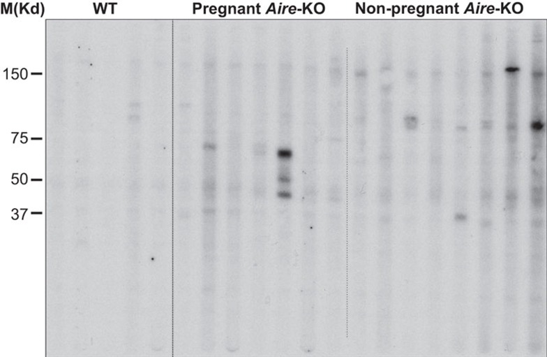
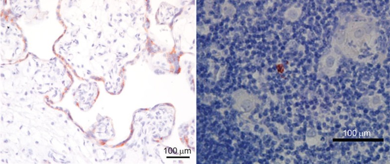
The ovary is not an immunologically privileged organ, but a breakdown in tolerogenic mechanisms for ovary-specific antigens has disastrous consequences on fertility in women, and this is replicated in murine models of autoimmune disease. Isolated ovarian autoimmune disease is rare in women, likely due to the severity of the disease and the inability to transmit genetic information conferring the ovarian disease across generations. Nonetheless, autoimmune oophoritis is often observed in association with other autoimmune diseases, particularly autoimmune adrenal disease, and takes a toll on both society and individual health. Studies in mice have revealed at least two mechanisms that protect the ovary from autoimmune attack. These mechanisms include control of autoreactive T cells by thymus-derived regulatory T cells, as well as a role for the autoimmune regulator (AIRE), a transcriptional regulator that induces expression of tissue-restricted antigens in medullary thymic epithelial cells during development of T cells. Although the latter mechanism is incompletely defined, it is well established that failure of either results in autoimmune-mediated targeting and depletion of ovarian follicles. In this review, we will address the clinical features and consequences of autoimmune-mediated ovarian infertility in women, as well as the possible mechanisms of disease as revealed by animal models.
Figures
Similar articles
-
The autoimmune regulator prevents premature reproductive senescence in female mice.Biol Reprod. 2012 Apr 12;86(4):110. doi: 10.1095/biolreprod.111.097501. Print 2012 Apr. Biol Reprod. 2012. PMID: 22219212 Free PMC article.
-
Autoimmune ovarian inflammation triggered by proinflammatory (Th1) T cells is compatible with normal ovarian function in mice.Biol Reprod. 1999 Sep;61(3):635-42. doi: 10.1095/biolreprod61.3.635. Biol Reprod. 1999. PMID: 10456839
-
[Autoimmune mechanisms of ovarian pathology].Fiziol Zh (1994). 2011;57(4):90-5. Fiziol Zh (1994). 2011. PMID: 22167839 Review. Ukrainian.
-
Autoimmune disease of the ovary induced by a ZP3 peptide from the mouse zona pellucida.J Clin Invest. 1992 Jan;89(1):28-35. doi: 10.1172/JCI115572. J Clin Invest. 1992. PMID: 1370297 Free PMC article.
-
Autoimmune disorders of the ovary.Indian J Exp Biol. 1998 May;36(5):433-6. Indian J Exp Biol. 1998. PMID: 9717459 Review.
Cited by
-
Protective role of stem cells in POI: Current status and mechanism of action, a review article.Heliyon. 2023 Dec 6;10(1):e23271. doi: 10.1016/j.heliyon.2023.e23271. eCollection 2024 Jan 15. Heliyon. 2023. PMID: 38169739 Free PMC article. Review.
-
Fetal-placental antigens and the maternal immune system: Reproductive immunology comes of age.Immunol Rev. 2022 Jul;308(1):25-39. doi: 10.1111/imr.13090. Epub 2022 May 29. Immunol Rev. 2022. PMID: 35643905 Free PMC article. Review.
-
The Role of T Cells in Ovarian Physiology and Infertility.Front Cell Dev Biol. 2022 Apr 26;10:713650. doi: 10.3389/fcell.2022.713650. eCollection 2022. Front Cell Dev Biol. 2022. PMID: 35557956 Free PMC article. Review.
-
Reproductive immunology issue 2: cellular and molecular biology.Cell Mol Immunol. 2014 Nov;11(6):503-5. doi: 10.1038/cmi.2014.98. Epub 2014 Sep 29. Cell Mol Immunol. 2014. PMID: 25263487 Free PMC article. No abstract available.
-
Human Amniotic Epithelial Stem Cells Alleviate Autoimmune Premature Ovarian Insufficiency in Mice by Targeting Granulosa Cells via AKT/ERK Pathways.Stem Cell Rev Rep. 2024 Aug;20(6):1618-1635. doi: 10.1007/s12015-024-10745-z. Epub 2024 Jun 4. Stem Cell Rev Rep. 2024. PMID: 38831179 Free PMC article.
References
-
- Forti G, Krausz C. Evaluation and treatment of the infertile couple. J Clin Endocrinol Metab. 1998;83:4177–4188. - PubMed
-
- Chandra A, Copen CE. Infertility and impaired fecundity in the United States, 1982–2010: data from the National Survey of Family Growth. Natl Health Stat Rep. 2013;67:1–19. - PubMed
-
- Braxton J, Carey D, Davis D, Footman A, Flagg E, Grier L, et al. Sexually Transmitted Disease Surveillance 2012. Atlanta, GA: Department of Health and Human Services: National Center for HIV/AIDS, Viral Hepatitis, STD, and TB Prevention; 2013.
-
- Brison DR, Roberts SA, Kimber SJ. How should we assess the safety of IVF technologies. Reprod Biomed Online. 2013;27:710–721. - PubMed
Publication types
MeSH terms
Substances
Grants and funding
LinkOut - more resources
Full Text Sources
Other Literature Sources
Medical
