Review
. 2014;78(11):2569-76.
doi: 10.1253/circj.cj-14-1080. Epub 2014 Oct 20.

Insulin regulation of myocardial autophagy

Affiliations
Review

Insulin regulation of myocardial autophagy

Christian Riehle et al. Circ J. 2014.

Abstract

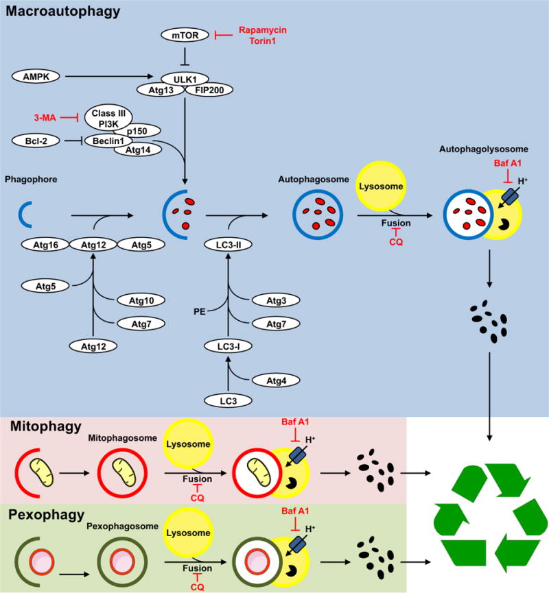
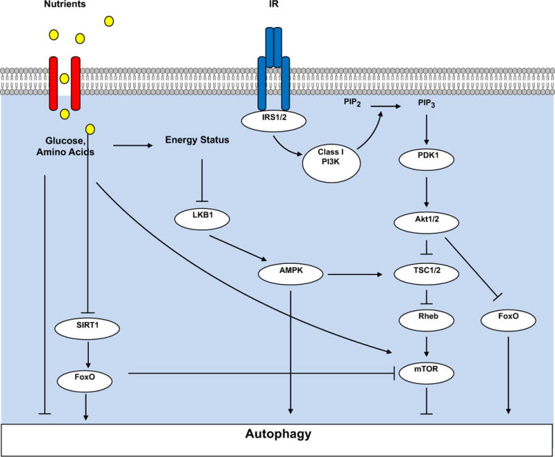
Autophagy is a conserved cellular process that plays an important role in cardiovascular homeostasis. Basal levels of autophagy are required for the maintenance of organellar quality control. Autophagy is dynamically regulated in the heart in the fasting to re-feeding transition. Insulin signaling plays an important role in the regulation of myocardial fuel metabolism, mitochondrial function and cellular growth. Recent studies have suggested an important role for insulin signaling in the regulation of myocardial autophagy. This dynamic regulation of autophagy induction during fasting may contribute to organellar homeostasis and if perturbed under conditions of hyperinsulinemia could contribute to accelerated cardiac aging.


Conflict of interest statement

Conflict of Interest Statement

The authors have no conflicts of interest to declare.

Figures

Figure 1
Figure 1. Schematic Representation of Insulin Signaling Pathways
Insulin receptors (IR) and Insulin Like Growth Factor 1 receptors (IGF1R) are tyrosine kinase cell surface receptors that exist as homodimers or as hybrid heterodimers. Activation of these receptors leads to association with insulin receptor substrate isoforms (IRS1/2), which are signaling scaffolds that facilitate activation of intracellular signaling molecules such as phosphoinositide-3-kinase (PI3K), which converts phosphatidylinositol (3,4)-bisphosphate (PIP2) to phosphatidylinositol (3,4,5)-trisphosphate (PIP3) at the plasma membrane that interacts with the PH-domain containing protein Phosphoinositide Dependent Kinase (PDK1) that initiates activating phosphorylation of the serine threonine kinase Akt. Akt phosphorylates many intracellular targets that include: the Forkhead box O (FOXO1) transcription factor leading to its nuclear exclusion to reduce its transcriptional activity; tuberous sclerosis complex 2 (TSC2) which reverses its repression of Rheb leading to activation of mechanistic target of rapamycin (mTOR); Glycogen synthase kinase 3 beta (GSK3b) removing the repression of glycogen synthase thereby stimulating glycogen synthesis and by phosphorylating AS160 (not shown) leading to translocation of GLUT4 glucose transporters to increase glucose uptake. IRS activation also increases phosphorylation of the Mitogen Activated Protein Kinases (ERK1 and 2) that regulate gene expression. Insulin Receptors and IRS proteins also forms complexes with other receptor families as exemplified by beta-adrenergic receptors to modulate ERK and PI3K signaling.
Figure 2
Figure 2. Summary of Signaling Mediators of Autophagy
A family of autophagy regulating proteins (Atg) are involved in specific steps in autophagy including biogenesis of autophagosome membranes, membrane expansion and genesis of autophagosomes and sequestration of cargo. Reagents in red represent pharmacological inhibitors that are commonly used to block specific autophagic steps. 3-MA – 3 methyl adenine; CQ – Chloroquine; BafA – Bafilomycin A. The insets provide illustrations of examples of two organelle-specific pathways of autophagy, namely mitophagy and pexophagy in which mitochondria and peroxisomes respectively, are specifically targeted to autophagosomes. Signaling molecules that regulate mitophagy are summarized in the text. Once formed autophagosomes fuse with lysosomes and their content is digested and recycled.
Figure 3
Figure 3. Regulation of Autophagy by Nutrient Status
Nutrients such as amino acids or glucose can directly activate mTOR, which represses autophagy. Under conditions of nutrient deficiency, activation of AMP activated protein kinase (AMPK) by binding to AMP and promoting its phosphorylation by Liver kinase B1 (LKB1). Activated AMPK will phosphorylate and inhibit mTOR leading to autophagy activation. AMPK may also directly activate autophagy by phosphorylating the autophagy mediator unc-51 like autophagy activating kinase 1 (ULK1)(not shown). Nutrient deficiency increases NAD+, which activates the NAD –dependent deactylase Sirtuin 1 (SIRT1), which will de-acetylate FOXO1, which is preferentially retained in the nucleus to increase its transcriptional activity on the promoters of genes encoding proteins that regulate autophagy. Increased nutrient availability promotes insulin release and activation of insulin receptors, which as summarized in Figure 1 leads to activation of mTOR, which suppresses autophagy and to phosphorylation of FOXO1, which excludes it from the nucleus, to lower its transcriptional activity on the promoters of genes that regulate autophagy.

References

    1. Muniyappa R, Montagnani M, Koh KK, Quon MJ. Cardiovascular actions of insulin. Endocr Rev. 2007;28:463–491. - PubMed
    1. Abel ED, O’Shea KM, Ramasamy R. Insulin resistance: metabolic mechanisms and consequences in the heart. Arterioscler Thromb Vasc Biol. 2012;32:2068–2076. - PMC - PubMed
    1. Wende AR, Symons JD, Abel ED. Mechanisms of lipotoxicity in the cardiovascular system. Curr Hypertens Rep. 2012;14:517–531. - PMC - PubMed
    1. Symons JD, McMillin SL, Riehle C, Tanner J, Palionyte M, Hillas E, et al. Contribution of insulin and Akt1 signaling to endothelial nitric oxide synthase in the regulation of endothelial function and blood pressure. Circ Res. 2009;104:1085–1094. - PMC - PubMed
    1. Rask-Madsen C, King GL. Vascular complications of diabetes: mechanisms of injury and protective factors. Cell Metab. 2013;17:20–33. - PMC - PubMed

Publication types