

Expression of Ki-67 in normal oral epithelium, leukoplakic oral epithelium and oral squamous cell carcinoma
- PMID: 25328294
- PMCID: PMC4196282
- DOI: 10.4103/0973-029X.140729
Expression of Ki-67 in normal oral epithelium, leukoplakic oral epithelium and oral squamous cell carcinoma
Abstract
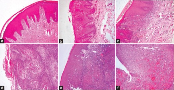
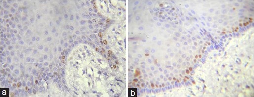
Aims and objective: To demonstrate the presence, location and pattern of cell proliferation in different histological grades of oral epithelial dysplasia (OED), oral squamous cell carcinoma (OSCC) and normal oral epithelium (NOE) using an antibody directed against the Ki-67 antigen and its intensity of staining evaluated respectively.
Materials and methods: A total number of 100 archival paraffin embedded blocks obtained from Department of Oral and Maxillofacial Pathology were studied. The case details were retrieved which consisted of histopathologically diagnosed cases of OSCC (n = 20), low risk OED (n = 30), high risk OED (n = 30) and normal appearing mucosa (n = 20) were taken as standard for comparison. Ki-67 immunostaining was detected. Ki-67 positive cells were counted in the five random high power fields in each case.
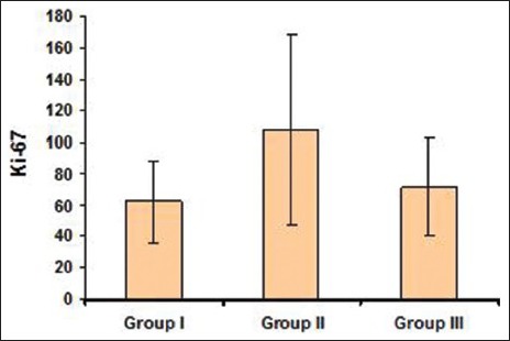
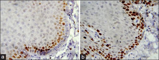
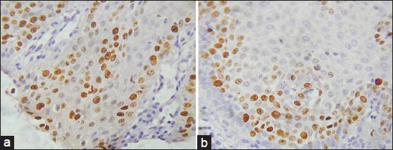
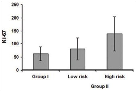
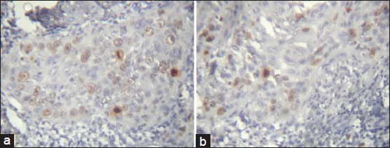
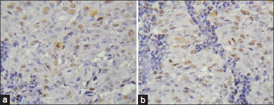
Results: Ki-67 labeling Index (LI) was restricted to the basal and parabasal layers of the normal oral epithelium irrespective of age, sex and site whereas it was seen in the basal, suprabasal and spinous layers in OED. Ki-67 LI is increased in high risk cases than the low risk cases of OED. Ki-67 positive cells in OSCC were located in the periphery of the tumor nests than the center, where frequent mitoses were observed.
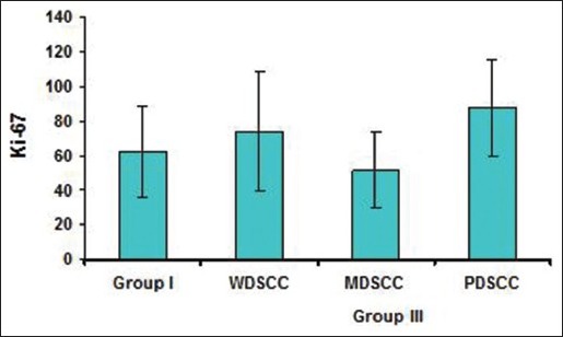
Conclusion: The architectural alteration evaluated by Ki-67 antibody in proliferating cell distribution in the layers of epithelial dysplasias may provide useful information to evaluate the grading of OED. Ki-67 LI increased in high risk cases than low risk cases of OED. This study showed that over expression of Ki-67 antigen between well-differentiated and poorly differentiated OSCC was in accordance with histologic grade of malignancy but not in accordance with moderately differentiated OSCC.
Keywords: Cell proliferation; Ki-67 LI (Labeling Index); Oral epithelial dysplasia; Oral squamous cell carcinoma.
Conflict of interest statement
Figures
References
-
- David H Cormach. Textbook of Ham's Histology. In: Barnes D, Winters R, Maxwell ME, editors. 9th ed. Philadelphia: J. B Lippincott Company; 1987. pp. 1–23.
-
- Pardee AB. G1 events and regulation of cell proliferation. Science. 1989;246:603–8. - PubMed
-
- Tumuluri V, Thomas GA, Fraser IS. Analysis of the Ki-67 antigen at the invasive tumour front of human oral squamous cell carcinoma. J Oral Pathol Med. 2002;31:598–604. - PubMed
-
- Bacci CE, Gown AM. Detection of cell proliferation in tissue sections. Braz J Med Biol Res. 1993;26:677–87. - PubMed
-
- Hahn WC, Weinberg RA. Rules for making human tumor cells. N Engl J Med. 2002;347:1593–603. - PubMed
LinkOut - more resources
Full Text Sources
Other Literature Sources
