

Adenoid cystic carcinoma: An unusual presentation
- PMID: 25328314
- PMCID: PMC4196302
- DOI: 10.4103/0973-029X.140796
Adenoid cystic carcinoma: An unusual presentation
Abstract
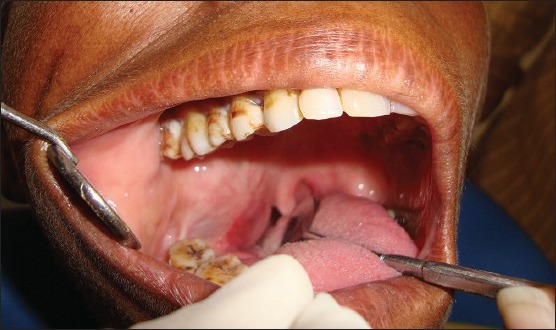
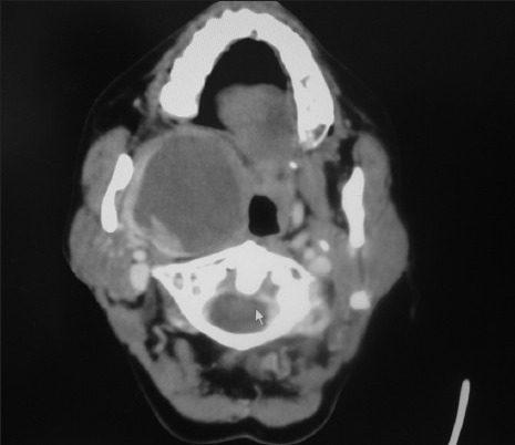
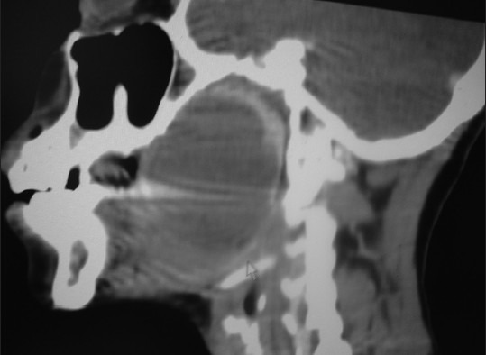
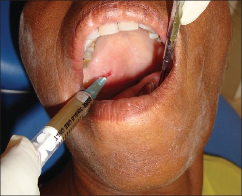
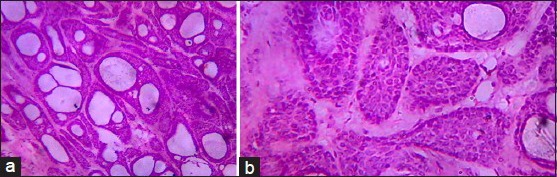
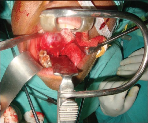
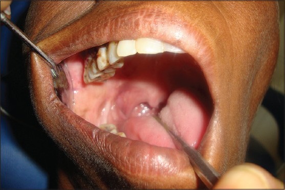
The adenoid cystic carcinoma is a relatively rare epithelial tumor of the major and minor salivary glands, accounting for about 1% of all malignant tumor of the oral and maxillofacial regions. Peak incidence occurs between the 5(th) and 6(th) decades of life. The clinical and pathological findings typical of this tumor include slow growth, peri-neural invasion, multiple local recurrences and distant metastasis. Herein, we report a case of adenoid cystic carcinoma of oropharynx with unusual clinical presentation. The diagnosis of this case and importance of cytology in diagnosing such cases is discussed.
Keywords: Adenoid cystic carcinoma; cribriform pattern; salivary gland malignancy.
Conflict of interest statement
Figures
Similar articles
-
Adenoid cystic carcinoma: a rare clinical entity and literature review.Oral Oncol. 2011 Apr;47(4):231-6. doi: 10.1016/j.oraloncology.2011.01.009. Epub 2011 Feb 24. Oral Oncol. 2011. PMID: 21353624 Review.
-
Adenoid cystic carcinoma located on the lower lip.Dermatol Online J. 2021 Sep 15;27(9). doi: 10.5070/D327955142. Dermatol Online J. 2021. PMID: 34755984
-
Adenoid cystic carcinoma of the parotid gland.Contemp Clin Dent. 2012 Apr;3(2):223-6. doi: 10.4103/0976-237X.96838. Contemp Clin Dent. 2012. PMID: 22919230 Free PMC article.
-
An unusual presentation of adenoid cystic carcinoma of the minor salivary glands with cranial nerve palsy: a case study.BMC Cancer. 2007 Aug 13;7:157. doi: 10.1186/1471-2407-7-157. BMC Cancer. 2007. PMID: 17697321 Free PMC article.
-
A rare case of adenoid cystic carcinoma of the nasopharynx manifesting as Horner's syndrome: discussion and review of the literature.Acta Otorhinolaryngol Ital. 2007 Aug;27(4):216-9. Acta Otorhinolaryngol Ital. 2007. PMID: 17957854 Free PMC article. Review.
Cited by
-
Microscopic Coblator Assisted Excision of Adenocystic Carcinoma of Soft Palate: A Novel Modality.Indian J Otolaryngol Head Neck Surg. 2025 Feb;77(2):652-658. doi: 10.1007/s12070-024-05203-8. Epub 2024 Nov 20. Indian J Otolaryngol Head Neck Surg. 2025. PMID: 40070762
-
Unusual presentation of adenoid cystic carcinoma (ACC) on lip mimicking mucocele: A rare case report with review.J Oral Maxillofac Pathol. 2023 Jul-Sep;27(3):605. doi: 10.4103/jomfp.jomfp_479_22. Epub 2023 Sep 12. J Oral Maxillofac Pathol. 2023. PMID: 38033939 Free PMC article.
-
Posterior fossa giant adenoid cystic carcinoma with skull base invasion mimicking glomus jugulare: A case report and review of literature.Rare Tumors. 2023 Jan 6;15:20363613221150218. doi: 10.1177/20363613221150218. eCollection 2023. Rare Tumors. 2023. PMID: 36636105 Free PMC article.
-
Sinonasal adenoid cystic carcinoma-role of on-site FNAC: a case report.BMC Ear Nose Throat Disord. 2018 May 9;18:6. doi: 10.1186/s12901-018-0053-4. eCollection 2018. BMC Ear Nose Throat Disord. 2018. PMID: 29760580 Free PMC article.
-
Current landscape and future directions of therapeutic approaches for adenoid cystic carcinoma of the salivary glands (Review).Oncol Lett. 2025 Jan 22;29(3):153. doi: 10.3892/ol.2025.14899. eCollection 2025 Mar. Oncol Lett. 2025. PMID: 39898287 Free PMC article. Review.
References
-
- Szanto PA, Luna MA, Tortoledo ME, White RA. Histologic grading of adenoid cystic carcinoma of the salivary glands. Cancer. 1984;54:1062–9. - PubMed
-
- Chummun S, McLean NR, Kelly CG, Dawes PJ, Meikle D, Fellows S, et al. Adenoid cystic carcinoma of the head and neck. Br J Plast Surg. 2001;54:476–80. - PubMed
-
- Nascimento AG, Amaral AL, Prado LA, Kligerman J, Silveira TR. Adenoid cystic carcinoma of salivary glands. A study of 61 cases with clinicopathologic correlation. Cancer. 1986;57:312–9. - PubMed
-
- Mendenhall WM, Morris CG, Amdur RJ, Werning JW, Hinerman RW, Villaret DB. Radiotherapy alone or combined with surgery for adenoid cystic carcinoma of the head and neck. Head Neck. 2004;26:154–62. - PubMed
-
- Spiro RH, Huvos AG, Strong EW. Adenoid cystic carcinoma: Factors influencing survival. Am J Surg. 1979;138:579–83. - PubMed
Publication types
LinkOut - more resources
Full Text Sources
Other Literature Sources
