. 2014:3:52-60.
doi: 10.1016/j.trivac.2014.02.003.

PROPHYLACTIC ANTIBODY TREATMENT AND INTRAMUSCULAR IMMUNIZATION REDUCE INFECTIOUS HUMAN RHINOVIRUS 16 LOAD IN THE LOWER RESPIRATORY TRACT OF CHALLENGED COTTON RATS

Affiliations

PROPHYLACTIC ANTIBODY TREATMENT AND INTRAMUSCULAR IMMUNIZATION REDUCE INFECTIOUS HUMAN RHINOVIRUS 16 LOAD IN THE LOWER RESPIRATORY TRACT OF CHALLENGED COTTON RATS

Jorge C G Blanco et al. Trials Vaccinol. 2014.

Abstract

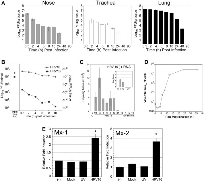
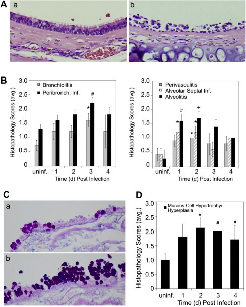
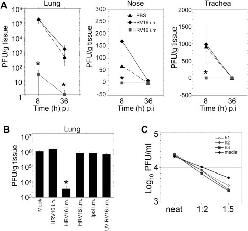
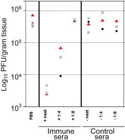
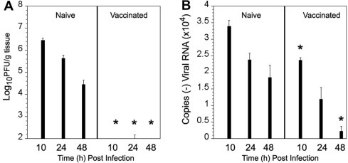
Human rhinoviruses (HRV) represent the single most important etiological agents of the common cold and are the most frequent cause of acute respiratory infections in humans. Currently the performance of available animal models for immunization studies using HRV challenge is very limited. The cotton rat (Sigmodon hispidus) is a well-recognized model for the study of human respiratory viral infections. In this work we show that, without requiring any genetic modification of either the host or the virus, intranasal infection of cotton rats with HRV16 resulted in measurable lower respiratory tract pathology, mucus production, and expression of interferon-activated genes. Intramuscular immunization with live HRV16 generated robust protective immunity that correlated with high serum levels of neutralizing antibodies. In addition, cotton rats treated prophylactically with hyperimmune anti-HRV16 serum were protected against HRV16 intranasal challenge. Finally, protection by immunization was efficiently transferred from mothers to newborn animals resulting in a substantial reduction of infectious virus loads in the lung following intranasal challenge. Overall, our results demonstrate that the cotton rat provides valuable additional model development options for testing vaccines and prophylactic therapies against rhinovirus infection.


Figures

Fig. 1
Fig. 1
Cotton rat infection with HRV16. Cotton rats were infected i.n. with 107 PFU of HRV16. (A) Infectious virus titers in nose, trachea, and lung homogenates from infected animals at the indicated times (h) post-infection. Groups of 5–10 animals were euthanized at each time in a total of 3 independent experiments. (B) Lung viral loads in rats infected with HRV16 compared to those of rats infected with HRV1B. Left axis shows input virus. (C) Quantification of (−) vRNA by RT-PCR in lung tissue at the indicated times post-infection. Animals inoculated with UV-inactivated HRV16 were used as control. Insert is a blow-out of the 24 h and 48 h time points from HRV16-infected and UV-HRV16-inoculated animals using logarithmic scale (n = 5 per time point). (D) One-cycle replication of HRV16 in HeLa Ohio cells. (E) qPCR quantification of Mx-1 and Mx-2 transcripts in BAL cells from uninfected or mock-inoculated rats, and rats instilled with UV-inactivated HRV16 or live HRV16 at 6 h after challenge. n = 8 per group. p < 0.05 in Student t-test analysis between HRV16-infected group and each of the control groups.
Fig. 2
Fig. 2
Airway pathology in HRV16-infected cotton rats. (A) Tracheal epithelial cell defoliation in HRV16-infected animals. (a) Trachea from an uninfected, or (b) HRV16-infected cotton rat. H&E staining, magnification 100×. (B) Histopathology scores obtained from lungs of uninfected (uninf) or HRV16-infected animals and euthanized at the indicated days post-infection. Graphs represent the extent of peribronchiolar infiltration, bronchiolitis, and epithelial degeneration (left); perivascular infiltration, alveolar septal infiltration and alveolar exudates (right). n = 3–9. p < 0.05, #p < 0.01, +p < 0.005 in one way Kruskal–Wallis ANOVA. (C) Alcian blue-PAS staining of a bifurcation of the main axial airway of an uninfected (a), or a HRV16-infected animal (b). (D) Scores for mucous cell hypertrophy/hyperplasia. p < 0.05, #p < 0.01 in one way Kruskal–Wallis ANOVA.
Fig. 3
Fig. 3
Immunogenicity and efficacy of immunization with live HRV16. (A) Viral titers in the lung, nose, and trachea of animals challenged with HRV16 (107 PFUs) 21 days post i.m. injection of PBS, post i.n. infection with 107 PFUs of HRV16, or post i.m. immunization with 107 PFUs HRV16. n = 5 per group. p < 0.05 in Student t-test analysis between HRV16-i.n. immunized group and the PBS-injected control group. (B) Immunization with different viral preparations showed that protection by immunization was specific for HRV16 and the intramuscular route. (C) Neutralization of viral infection occurs in vivo. Lung homogenates from 3 animals previously immunized with HRV16 (neutralization titers > 1280), subsequently challenged and sacrificed 10 h after challenge (h1, h2, h3) were mixed with homogenates of a naïve, challenged animal (PBS i.m.) also sacrificed 10 h p.i. Mixed homogenates in the indicated dilution (immune:control) were then titrated for determination of viral load. A control curve (media) was performed by diluting a control homogenate with titration media. The reduction of resulting titers in all mixes was consistent with the dilution factor and not consistent with neutralization occurring ex vivo.
Fig. 4
Fig. 4
Passive HRV16 antibody transfer protects animals from HRV16 challenge. Animals were treated intraperitoneally with control cotton rat serum (anti-HRV16 neutralization titers < 20) or with different serum dilutions from animals immunized i.m. with HRV16 (1 × 106 PFU). At 1 day post-treatment, animals were challenged i.n. with 107 PFUs of HRV16, and euthanized 8 h later to determine lung viral titers. Each symbol corresponds to one animal.
Fig. 5
Fig. 5
Maternal immunity confers protection to pups. (A) Newborns from naïve or HRV16-immunized females were challenged i.n. with ∼5 × 106 PFUs of HRV16 in 20 μl. Infectious virus titers in the lung were determined by plaque assay at the indicated times p.i. (B) HRV16 (−) viral RNA detection in lungs of newborn cotton rats. n = 6–8 animals/group where each group consisted of pups from 2 different mothers. p < 0.05 in Student t-test comparison between group of pups whose mothers were immunized vs groups of pups of the same age from unimmunized, naïve mothers.

Similar articles

Cited by

References

    1. Arruda E., Pitkaranta A., Witek T.J., Jr., Doyle C.A., Hayden F.G. Frequency and natural history of rhinovirus infections in adults during autumn. J. Clin. Microbiol. 1997;35:2864–2868. - PMC - PubMed
    1. Gern J.E., Palmenberg A.C. Rhinoviruses. Chapter 18. In: Knipe D.M., Howley P.M., editors. sixth ed. vol. 1. Lippincott-Williams & Wilkins; Philadelphia: 2013. p. 531. (Fields Virology).
    1. Jacobs S.E., Lamson D.M., St George K., Walsh T.J. Human rhinoviruses. Clin. Microbiol. Rev. 2013;26:135–162. - PMC - PubMed
    1. Knowles N.J., Hovi T., Hyypiä T., King A.M.Q., Lindberg A.M. Picornaviridae. In: King A.M.Q., Adams M.J., Carstens E.B., Lefkowitz E.J., editors. Virus Taxonomy: Classification and Nomenclature of Viruses: Ninth Report of the International Committee on Taxonomy of Viruses. Elsevier; San Diego: 2012. pp. 855–880.
    1. McIntyre C.L., Knowles N.J., Simmonds P. Proposals for the classification of human rhinovirus species A, B and C into genotypically assigned types. J. Gen. Virol. 2013;94:1791–1806. - PMC - PubMed