

Novel technique for innervated abdominal wall vascularized composite allotransplantation: a separation of components approach
- PMID: 25328567
- PMCID: PMC4171836
Novel technique for innervated abdominal wall vascularized composite allotransplantation: a separation of components approach
Abstract
Objective: Applications for Abdominal Wall Vascularized Composite Allotransplantation may expand if a functional graft with decreased immunosuppressive requirements can be designed. We hypothesize that it is anatomically feasible to prepare a functional, innervated, and vascularized abdominal composite graft using a multilayered component separation technique. Including vascularized bone in the graft design may decrease the immunosuppressive requirements by inducing immunologic chimerism.
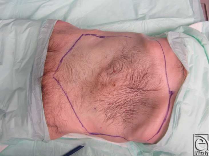
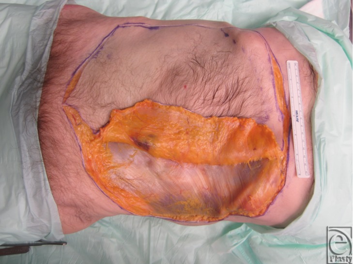
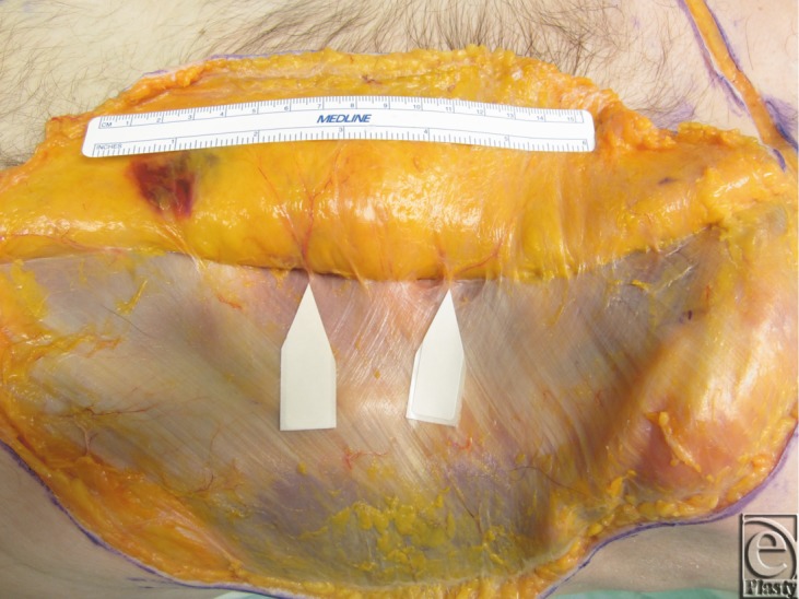
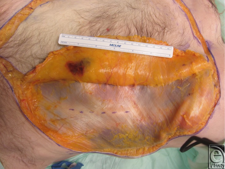
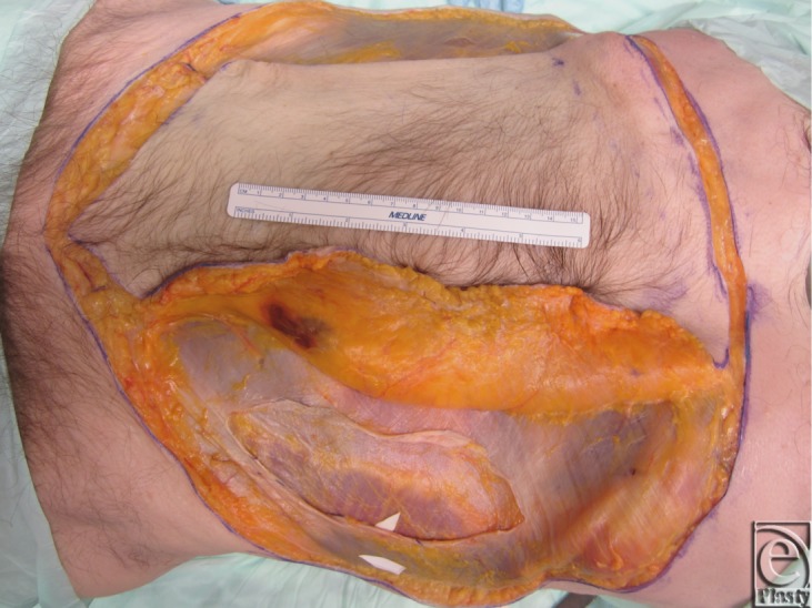
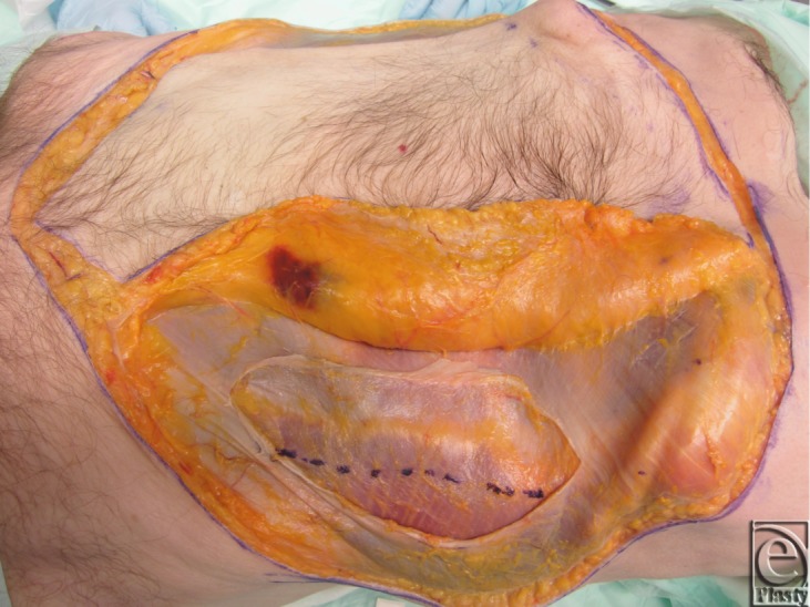
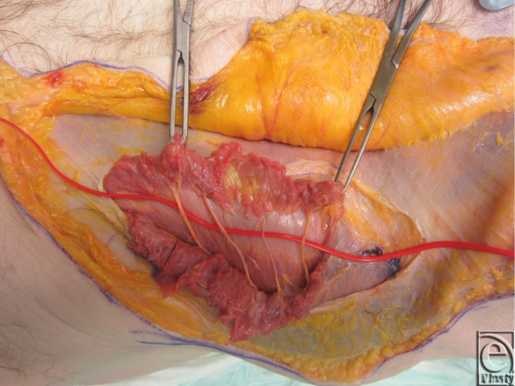
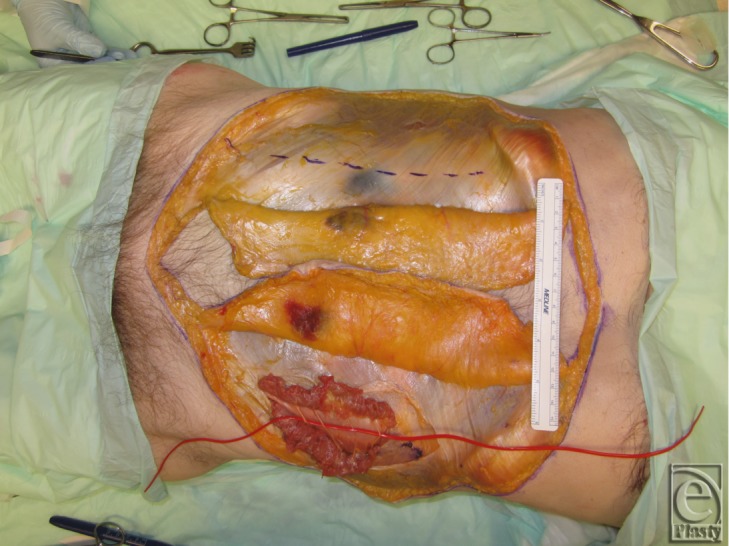
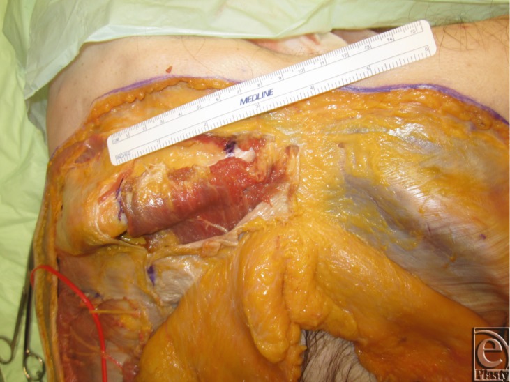
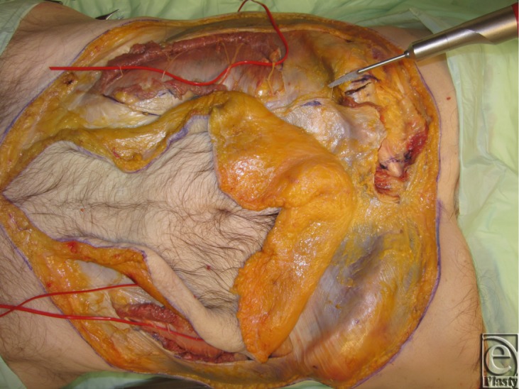
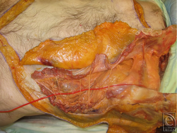
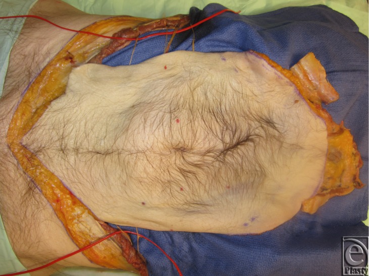
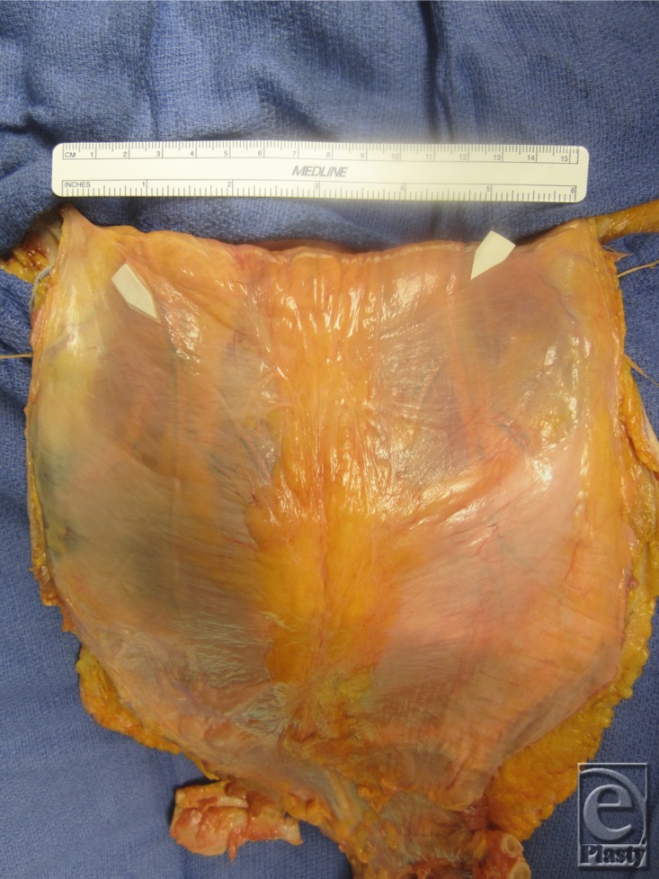
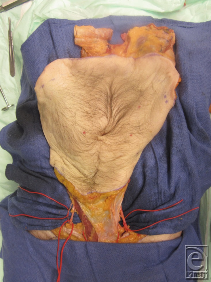
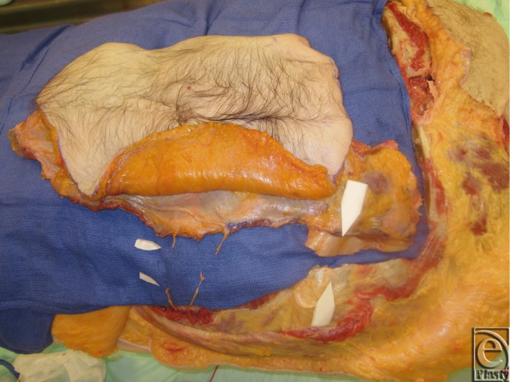
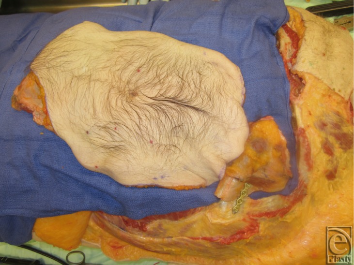
Methods: Two cadaver torsos were used. Adipocutaneous flaps were elevated from the midaxillary lines, preserving deep inferior epigastric artery perforators. A 2-layered component separation through the external and internal oblique fasciae was carried out, exposing segmental intercostal thoracolumbar nerves. Superiorly directed muscle release over the subcostal margin provided for a 3-rib segment with attached rectus abdominis muscle. The remainder of the full-thickness allograft was harvested with its vasculature. Flap inset into the recipient cadaver abdomen, with osteosynthesis fixation between donor and recipient ribs, was achieved.
Results: The harvested grafts had an average size of 845 ± 205 cm(2) with a total procurement time of 110 minutes. On one cadaver, 4 thoracolumbar nerves were isolated bilaterally, while the other cadaver yielded 3 nerves. The nerves were transected with an average length of 5.7 ± 1.2 cm. The graft vasculature was transected with a length of 4.40 ± 0.10 cm.
Conclusion: Using the principles of component separation technique, we demonstrated a novel approach to harvest and transfer a neurotized osteomyofasciocutaneous abdominal wall allotransplant as a multipedicled, single functional unit.
Keywords: abdominal wall transplantation; chimerism; component separation; hernia; vascularized composite allotransplantation.
Figures
References
-
- Azari KK, Imbriglia JE, Goitz RJ, et al. Technical aspects of the recipient operation in hand transplantation. J Reconstr Microsurg. 2012;28:27–34. - PubMed
-
- Dorafshar AH, Bojovic B, Christy MR, et al. Total face, double jaw, and tongue transplantation: an evolutionary concept. Plast Reconstr Surg. 2013;131:241–51. - PubMed
-
- Cipriani R, Contedini F, Santoli M, et al. Abdominal wall transplantation with microsurgical technique. Am J Transplant. 2007;7:1304–7. - PubMed
-
- Selvaggi G, Levi DM, Kato T, et al. Expanded use of transplantation techniques: abdominal wall transplantation and intestinal autotransplantation. Transplant Proc. 2004;36:1561–3. - PubMed
-
- Selvaggi G, Levi DM, Cipriani R, Sgarzani R, Pinna AD, Tzakis AG. Abdominal wall transplantation: surgical and immunologic aspects. Transplant Proc. 2009;41:521–2. - PubMed
LinkOut - more resources
Full Text Sources