. 2014 Aug;56(2):108-13.
doi: 10.3340/jkns.2014.56.2.108. Epub 2014 Aug 31.

Surgical outcomes after traumatic vertebral fractures in patients with ankylosing spondylitis

Affiliations

Surgical outcomes after traumatic vertebral fractures in patients with ankylosing spondylitis

Seong-Bae An et al. J Korean Neurosurg Soc. 2014 Aug.

Abstract

Objective: Ankylosing spondylitis is an inflammatory rheumatic disease mainly affecting the axial skeleton. The rigid spine may secondarily develop osteoporosis, further increasing the risk of spinal fracture. In this study, we reviewed fractures in patients with ankylosing spondylitis that had been clinically diagnosed to better define the mechanism of injury, associated neurological deficit, predisposing factors, and management strategies.

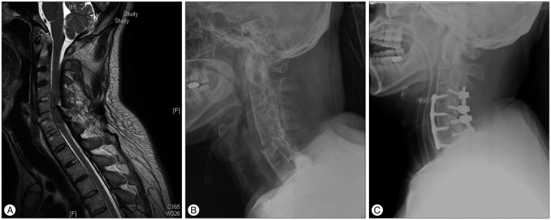
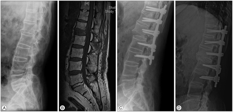
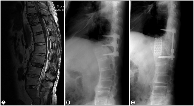
Methods: Between January 2003 and December 2013, 12 patients with 13 fractures with neurological complications were treated. Neuroimaging evaluation was obtained in all patients by using plain radiography, CT scan, and MR imaging. The ASIA Impairment Scale was used in order to evaluate the neurologic status of the patients. Management was based on the presence or absence of spinal instability.

Results: A total of 9 cervical and 4 thoracolumbar fractures were identified in a review of patients in whom ankylosing spondylitis had been diagnosed. Of these, 7 fractures were associated with a hyperextension mechanism. 10 cases resulted in a fracture by minor trauma. Posttraumatic neurological deficits were demonstrated in 11 cases and neurological improvement after surgery was observed in 5 of these cases.

Conclusions: Patients with ankylosing spondylitis are highly susceptible to spinal fracture and spinal cord injury even after only mild trauma. Initial CT or MR imaging of the whole spine is recommended even if the patient's symptoms are mild. The patient should also have early surgical stabilization to correct spinal deformity and avoid worsening of the patient's neurological status.

Keywords: Ankylosing spondylitis; Spinal cord injury; Surgery; Trauma; Vertebral fracture.


Figures

Fig. 1
Fig. 1
Case 5. Imaging study obtained in 38-year-old man suffering from ankylosing spondylitis for 15 years. The patient had been transferred to our center from a local hospital after falling down in a drunken state. When he arrived at our hospital, his neurologic status was quadriplegia (ASIA grade A). The patient underwent emergent two-stage anterior-posterior fixation to stabilization. After a 1-year follow-up period, his neurologic status was not improved and he suffered aspiration pneumonia. After a tracheostomy, he was transferred to a rehabilitation center. A and B : Preoperative MR imaging and plain radiography showing C5-6 instability, spinal cord injury due to compromised canal from the C6 lamina, and so-called bamboo spine resulting from ankylosing spondylitis. C : Plain radiography demonstrating the outcome after two-stage anterior-posterior stabilization.
Fig. 2
Fig. 2
Case 12. Imaging studies obtained in a 72-year-old man with known ankylosing spondylitis for 30 years. He experienced immediate bilateral leg motor weakness (ASIA grade C) from a fall-related injury at home. After surgery, the patient's neurological status was improved to ASIA grade E for 11 months of follow-up. A and B : Preoperative plain radiography and MR imaging reveal a flexion fracture and dislocation of L1. C : Immediate postoperative radiography after placement of pedicle screw from T11 to L3 and laminectomy L1, 2, 3. These figures show a structure of ankylosed spine like a long bone fracture and further displacement of the fracture site after surgery. It may have occurred in positioning the patient in the operating room. D : postoperative radiography for 11 months of follow-up demonstrating a fused state.
Fig. 3
Fig. 3
Case 10. A : Preoperative sagittal magnetic resonance T2-weighted image. Fracture had extended to the posterior column and the patient underwent pedicle screw fixation T10-11-12 and laminectomy T10, 11 at a local hospital. B : Plain radiography from when the patient arrived at our hospital after minor trauma. Fracture dislocation was noted because of implant failure. C : Plain radiography after 1 month of follow-up after operation in our hospital. Previous implants were removed; corpectomy and MESH cage implantation were performed by retroperitoneal extra-pleural approach.

Similar articles

Cited by

References

    1. Alaranta H, Luoto S, Konttinen YT. Traumatic spinal cord injury as a complication to ankylosing spondylitis. An extended report. Clin Exp Rheumatol. 2002;20:66–68. - PubMed
    1. Anwar F, Al-Khayer A, Joseph G, Fraser MH, Jigajinni MV, Allan DB. Delayed presentation and diagnosis of cervical spine injuries in long-standing ankylosing spondylitis. Eur Spine J. 2011;20:403–407. - PMC - PubMed
    1. Braun J, Sieper J. Ankylosing spondylitis. Lancet. 2007;369:1379–1390. - PubMed
    1. Caron T, Bransford R, Nguyen Q, Agel J, Chapman J, Bellabarba C. Spine fractures in patients with ankylosing spinal disorders. Spine (Phila Pa 1976) 2010;35:E458–E464. - PubMed
    1. Cornefjord M, Alemany M, Olerud C. Posterior fixation of subaxial cervical spine fractures in patients with ankylosing spondylitis. Eur Spine J. 2005;14:401–408. - PMC - PubMed

LinkOut - more resources