

Gallstone Ileus following Endoscopic Stone Extraction
- PMID: 25328725
- PMCID: PMC4195353
- DOI: 10.1155/2014/271571
Gallstone Ileus following Endoscopic Stone Extraction
Abstract
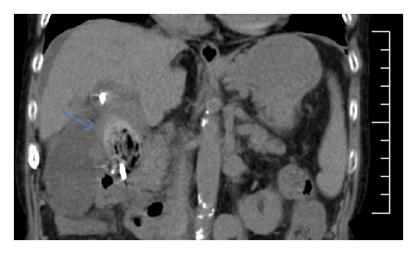
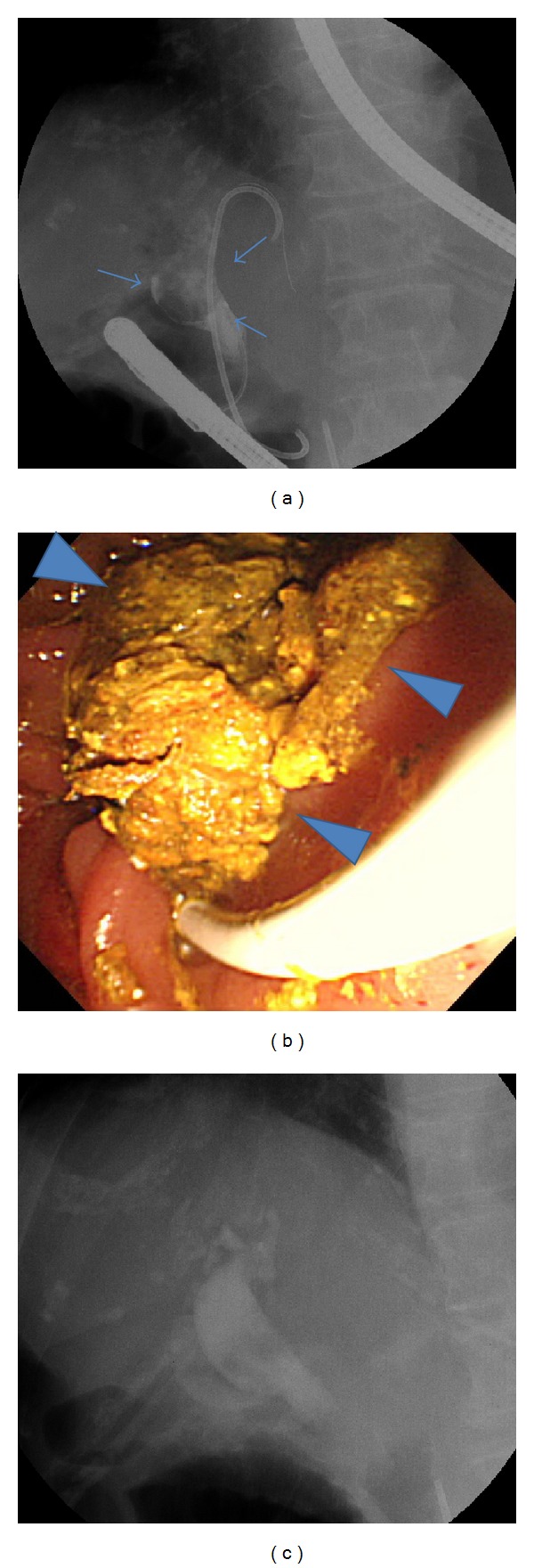
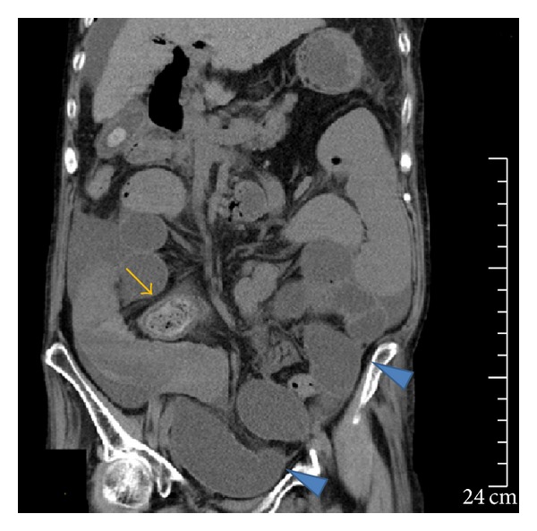
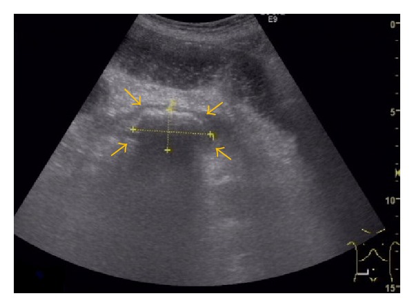
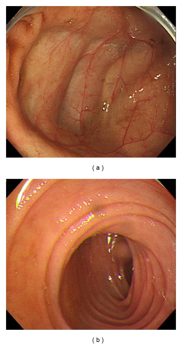
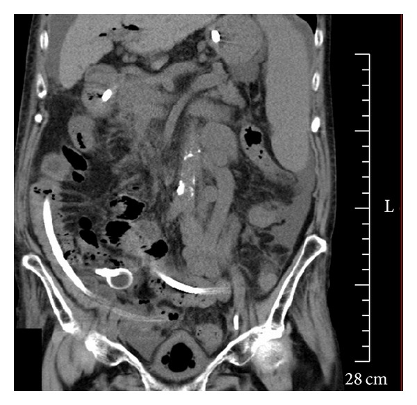
An 85-year-old woman was an outpatient treated at Tokyo Rosai Hospital for cirrhosis caused by hepatitis B. She had previously been diagnosed as having common bile duct stones, for which she underwent endoscopic retrograde cholangiopancreatography (ERCP). However, as stone removal was unsuccessful, a plastic stent was placed after endoscopic sphincterotomy. In October 2012, the stent was replaced endoscopically because she developed cholangitis due to stent occlusion. Seven days later, we performed ERCP to treat recurring cholangitis. During the procedure, the stone was successfully removed by a balloon catheter when cleaning the common bile duct. The next day, the patient developed abdominal pain, abdominal distension, and nausea and was diagnosed as having gallstone ileus based on abdominal computed tomography (CT) and abdominal ultrasonography findings of an incarcerated stone in the terminal ileum. Although colonoscopy was performed after inserting an ileus tube, no stone was visible. Subsequent CT imaging verified the disappearance of the incarcerated stone from the ileum, suggesting that the stone had been evacuated naturally via the transanal route. Although it is extremely rare for gallstone ileus to develop as a complication of ERCP, physicians should be aware of gallstone ileus and follow patients carefully, especially after removing huge stones.
Figures
References
-
- Reisner RM, Cohen JR. Gallstone ileus: a review of 1001 reported cases. The American Surgeon. 1994;60(6):441–446. - PubMed
-
- Hatano K, Ishihara Y, Noguchi T, Sonobe X. A case of gallstone ileus and review of cumulated 130 cases in the Japanese literature. The Journal of the Japanese Practical Surgeon Society. 1993;54(8):2150–2154.
-
- Halter F, Bangerter U, Gigon JP, Pusterla C. Gallstone ileus after endoscopic sphincterotomy. Endoscopy. 1981;13(2):88–89. - PubMed
-
- Oskam J, Heitbrink M, Schattenkerk ME. Intermittent gallstone ileus following endoscopic biliary sphincterotomy. A case report. Acta Chirurgica Belgica. 1993;93(2):43–45. - PubMed
LinkOut - more resources
Full Text Sources
Other Literature Sources
