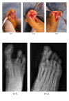
Osteoid osteoma of distal phalanx of toe: a rare cause of foot pain
- PMID: 25328736
- PMCID: PMC4190831
- DOI: 10.1155/2014/560892
Osteoid osteoma of distal phalanx of toe: a rare cause of foot pain
Abstract
Osteoid osteoma is an uncommon benign tumor and causes severe pain, being worse at night, that responds dramatically to nonsteroidal anti-inflammatory medications. An osteoid osteoma of the toe is very rare and arising in a pedal phalanx may be difficult to diagnose. A 34-year-old male has local swelling and tenderness but there were no hyperemia, temperature increase, or clubbing. There was a 2-month history of antibiotic treatment with suspicion of soft tissue infection in another clinic. The osteoid osteoma was completely excised by curettage and nidus removal with open surgical technique. The patient was followed up for 63 months with annual clinical and radiographic evaluations. There was no relapse of the pain and no residual recurrent tumour. Osteoid osteoma may be difficult to distinguish from chronic infection or myxedema. The patients may be taken for unnecessary treatment. The aim of the treatment for osteoid osteoma is to remove entire nidus by open surgical excision or by percutaneous procedures such as percutaneous radiofrequency and laser ablation. Osteoid osteomas having radiologic and clinical features other than classical presentation of osteoid osteoma are called atypical osteoid osteomas. Atypical localized osteoid osteomas can be easily misdiagnosed and treatment is often complicated.
Figures


Similar articles
-
Hallux Osteoid Osteoma: A Case Report and Literature Review.Open Orthop J. 2017 Sep 30;11:1066-1072. doi: 10.2174/1874325001711011066. eCollection 2017. Open Orthop J. 2017. PMID: 29151998 Free PMC article. Review.
-
A review of literature: Mosaicoplasty as an alternative treatment for resection of patellar osteoid osteoma and cartilage reconstruction.J Orthop. 2018 May 17;15(3):768-771. doi: 10.1016/j.jor.2018.05.036. eCollection 2018 Sep. J Orthop. 2018. PMID: 29946202 Free PMC article. Review.
-
[RARE LOCALIZATION OF OSTEOID OSTEOMA--DISTAL PHALANX OF THE RING FINGER].Acta Med Croatica. 2016 Sep;70(3):191-5. Acta Med Croatica. 2016. PMID: 29064211 Croatian.
-
Osteoid osteoma of the great toe.Orthopedics. 2011 Aug 8;34(8):e432-5. doi: 10.3928/01477447-20110627-33. Orthopedics. 2011. PMID: 21815591
-
Osteoid Osteoma of the Distal Phalanx: A Rare Condition.Cureus. 2021 Oct 27;13(10):e19077. doi: 10.7759/cureus.19077. eCollection 2021 Oct. Cureus. 2021. PMID: 34824948 Free PMC article.
Cited by
-
Osteoid Osteoma of the Toe: A Rare Presentation with Diagnostic Challenges.J Belg Soc Radiol. 2023 Aug 29;107(1):64. doi: 10.5334/jbsr.3250. eCollection 2023. J Belg Soc Radiol. 2023. PMID: 37664521 Free PMC article.
-
[Difficulties in diagnosing an osteoid osteoma of the big toe : A case report with review of the literature].Orthopade. 2021 Sep;50(9):689-699. doi: 10.1007/s00132-021-04082-z. Epub 2021 Apr 8. Orthopade. 2021. PMID: 33830283 Free PMC article. Review. German.
-
Osteoid Osteoma: A Unique Presentation in a Child's Lesser Toe.Case Rep Orthop. 2021 Jul 27;2021:8876584. doi: 10.1155/2021/8876584. eCollection 2021. Case Rep Orthop. 2021. PMID: 34367707 Free PMC article.
-
Hallux Osteoid Osteoma: A Case Report and Literature Review.Open Orthop J. 2017 Sep 30;11:1066-1072. doi: 10.2174/1874325001711011066. eCollection 2017. Open Orthop J. 2017. PMID: 29151998 Free PMC article. Review.
References
-
- Barca F, Acciaro AL, Recchioni MD. Osteoid osteoma of the phalanx: enlargement of the toe—two case reports. Foot and Ankle International. 1998;19(6):388–393. - PubMed
-
- Laurence N, Epelman M, Markowitz RI, Jaimes C, Jaramillo D, Chauvin NA. Osteoid osteomas: a pain in the night diagnosis. Pediatric Radiology. 2012;42(12):1490–1501. - PubMed
-
- Sproule JA, Khan F, Fogarty EE. Osteoid osteoma: painful enlargement of the second toe. Archives of Orthopaedic and Trauma Surgery. 2004;124(5):354–356. - PubMed
-
- McGrath BE, Bush CH, Nelson TE, Scarborough MT. Evaluation of suspected osteoid osteoma. Clinical Orthopaedics and Related Research. 1996;(327):247–252. - PubMed
-
- Meng Q, Watt I. Phalangeal osteoid osteoma. British Journal of Radiology. 1989;62(736):321–325. - PubMed
LinkOut - more resources
Full Text Sources
Other Literature Sources

