

Molecular Genetics of Mycobacteriophages
Affiliations
- PMID: 25328854
- PMCID: PMC4199240
Item in Clipboard
Molecular Genetics of Mycobacteriophages
Microbiol Spectr.
.
Abstract
Mycobacteriophages have provided numerous essential tools for mycobacterial genetics, including delivery systems for transposons, reporter genes, and allelic exchange substrates, and components for plasmid vectors and mutagenesis. Their genetically diverse genomes also reveal insights into the broader nature of the phage population and the evolutionary mechanisms that give rise to it. The substantial advances in our understanding of the biology of mycobacteriophages including a large collection of completely sequenced genomes indicates a rich potential for further contributions in tuberculosis genetics and beyond.
Figures
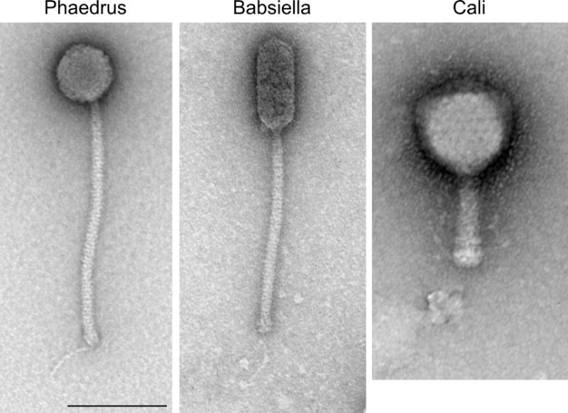
Mycobacteriophage morphologies. Three examples of virion morphologies are illustrated. Phaedrus and Babsiella exhibit siphoviral morphologies with long flexible tails; Phaedrus has an isometric head, whereas the Babsiella head is prolate. Cali is an example of myoviral morphology. Scale bar is 100 nm. 10.1128/microbiolspec.MGM2-0032-2013.f1
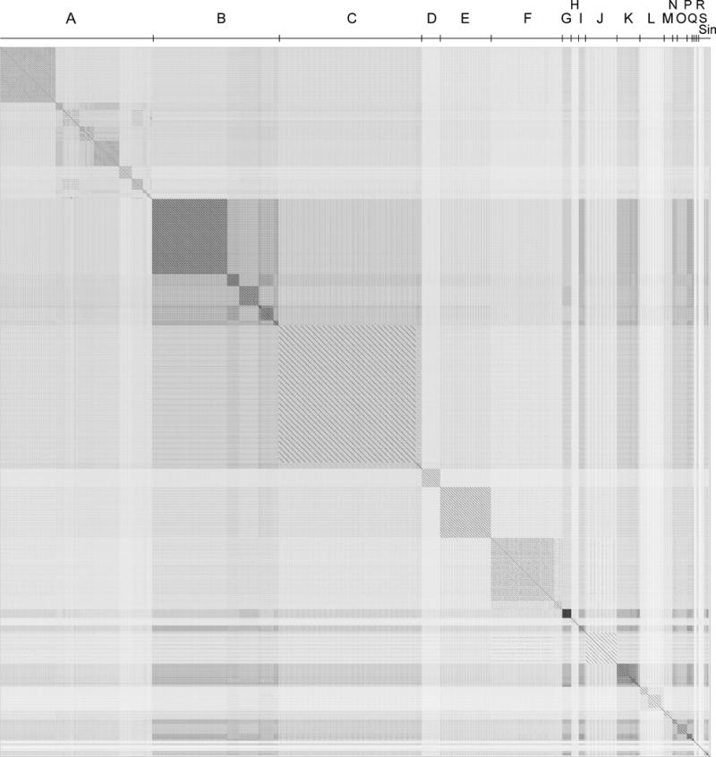
Dotplot comparison of 285 mycobacteriophage genomes. A concatenated file of 285 mycobacteriophage nucleotide sequences was compared against itself using the Gepard program (242) to generate the dotplot. The order of the genomes was arranged such that genomically related phages were adjacent to each other in this file, and the clusters of related phages (Clusters A, B, C, etc.) are shown above the plot. Five of the genomes are singletons with no closely related phages and are denoted collectively as Sin. 10.1128/microbiolspec.MGM2-0032-2013.f2
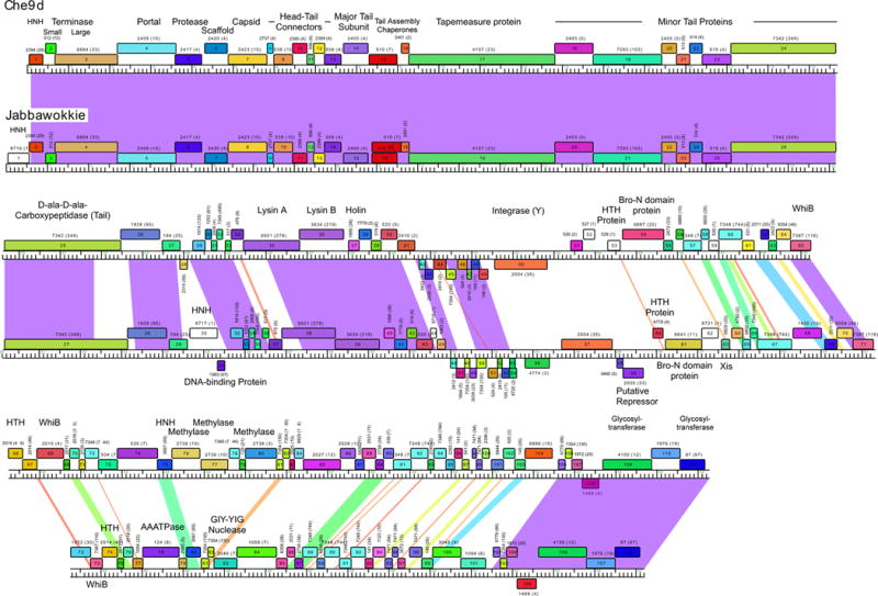
Comparison of mycobacteriophage Che9d and Jabbawokkie genome maps. Mycobacteriophages Che9d and Jabbawokkie are grouped into Subcluster F2, and their genome maps are shown as represented by the Phamerator program (100). Each genome is shown with markers, and the shading between the genomes reflects nucleotide sequence similarity determined by BLASTN, spectrum-colored with the greatest similarity in purple and the least in red. Protein-coding genes are shown as colored boxes above or below the genomes, reflecting rightward or leftward transcription, respectively. Each gene is assigned a phamily (Pham) designation based on amino acid sequence similarity (see text), as shown above or below each box, with the number of phamily members shown in parentheses; genes shown as white boxes are orphams and have no other phamily members. Putative gene functions are indicated. 10.1128/microbiolspec.MGM2-0032-2013.f3
Functional genomics of mycobacteriophage Giles. A map of the mycobacteriophage Giles was generated using Phamerator and annotated as described for Fig. 3. Boxes below the genome indicate whether the gene is nonessential for lytic growth (yellow), likely essential (blue), or essential (green). Arrows indicate genes expressed in lysogeny (red) or early (green) or late (purple) lytic growth, with line thickness reflecting transcription strength. Reproduced with permission from Dedrick et al. (107). 10.1128/microbiolspec.MGM2-0032-2013.f4
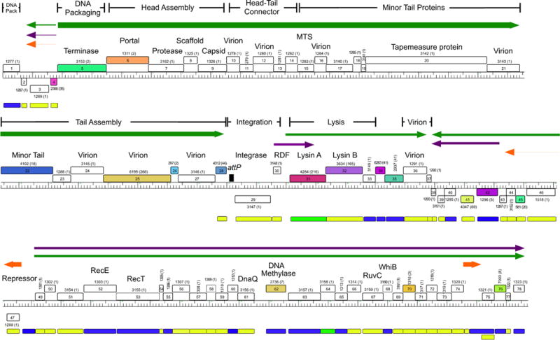
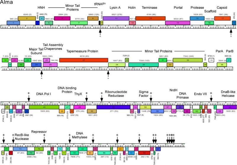
Genome map of mycobacteriophage Alma. The genome map of mycobacteriophage Alma was generated using Phamerator and is illustrated as described for Fig. 3. Alma is a Subcluster A6 phage and shares the features of other Cluster A phages in having multiple binding sites for its repressor protein (gp75). These stoperator sites are indicated by vertical arrows, and the orientation of the asymmetric sites relative to genome orientation are shown as (−) or (+). Stoperators were identified as sequences corresponding to the consensus sequence 5′-GATGAGTGTCAAG with no more than a single mismatch. Note that the stoperator consensus sequences can differ for different Subcluster A phages (98). 10.1128/microbiolspec.MGM2-0032-2013.f5
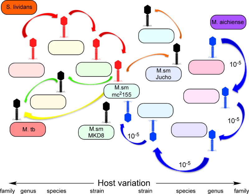
A model for mycobacteriophage diversity. The large number of different types of mycobacteriophages isolated on M. smegmatis mc2155 can be explained by a model in which phages can readily infect new bacterial hosts—either by a switch or an expansion of host range—using a highly diverse bacterial population that includes many closely related strains. As such, phages with distinctly different genome sequences and GC% contents infecting distantly related bacterial hosts, such as those to the left (red) or right (blue) extremes of a spectrum of hosts, can migrate across a microbial landscape through multiple steps. Each host switch occurs at a relatively high frequency (~1 in 105 particles, or an average of about one every 103 bursts of lytic growth) and much faster than either amelioration of phage GC% to its new host or genetic recombination. Two phages (such as those shown in red and blue) can thus “arrive” at a common host (M. smegmatis mc2155) but be of distinctly different types (clusters, subclusters, and singletons). Reproduced with permission from Jacobs-Sera et al. (99). 10.1128/microbiolspec.MGM2-0032-2013.f6
References
-
- Gardner GM, Weiser RS. A bacteriophage for Mycobacterium smegmatis. Proc Soc Exp Biol Med. 1947;66:205–206. - PubMed
-
- Whittaker E. Two bacteriophages for Mycobacterium smegmatis. Can J Public Health. 1950;41:431–436. - PubMed
-
- Grange JM. Proceedings: bacteriophage typing of strains of Mycobacterium tuberculosis isolated in south-east England. J Med Microbiol. 1975;8:ix(2). - PubMed
-
- Grange JM. The genetics of mycobacteria and mycobacteriophages: a review. Tubercle. 1975;56:227–238. - PubMed
Grants and funding
LinkOut - more resources
Full Text Sources