Review
. 2014:2014:687608.
doi: 10.1155/2014/687608. Epub 2014 Aug 14.

The role of 18F-FDG PET/CT in large-vessel vasculitis: appropriateness of current classification criteria?

Affiliations
Review

The role of 18F-FDG PET/CT in large-vessel vasculitis: appropriateness of current classification criteria?

H Balink et al. Biomed Res Int. 2014.

Abstract

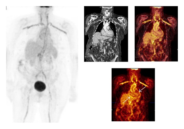
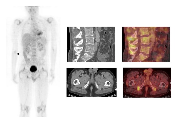
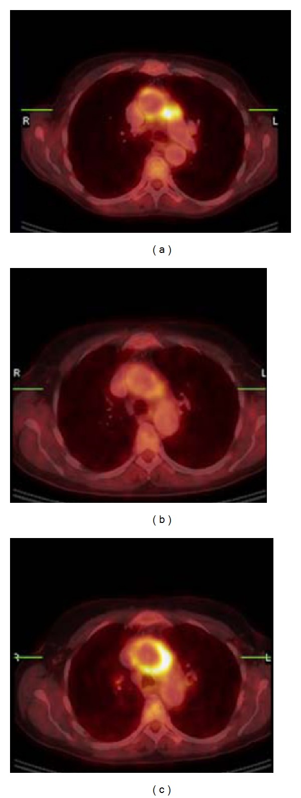
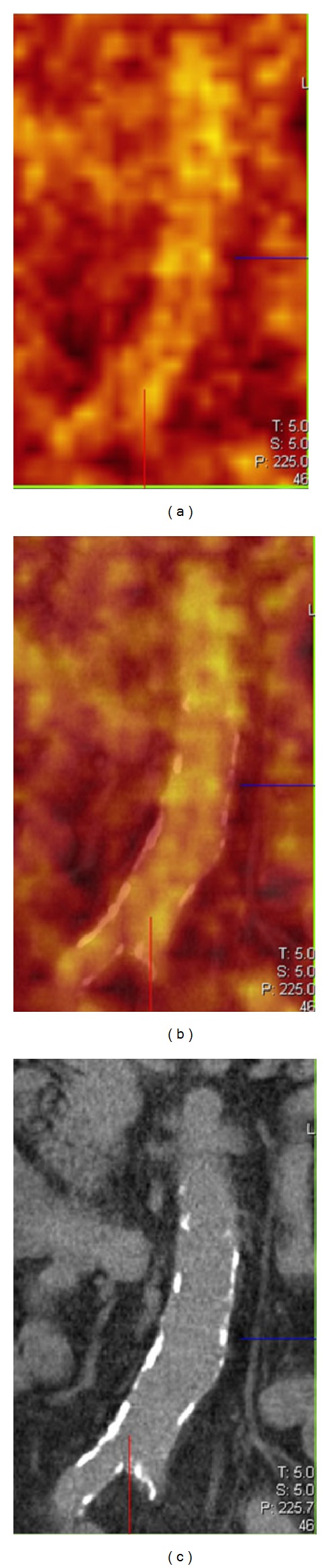
Patients with clinical suspicion of large-vessel vasculitis (LVV) may present with nonspecific signs and symptoms and increased inflammatory parameters and may remain without diagnosis after routine diagnostic procedures. Both the nonspecificity of the radiopharmaceutical (18)F-FDG and the synergy of integrating functional and anatomical images with PET/CT offer substantial benefit in the diagnostic work-up of patients with clinical suspicion for LVV. A negative temporal artery biopsy, an ultrasonography without an arterial halo, or a MRI without aortic wall thickening or oedema do not exclude the presence of LVV and should therefore not exclude the use of (18)F-FDG PET/CT when LVV is clinically suspected. This overview further discusses the notion that there is substantial underdiagnosis of LVV. Late diagnosis of LVV may lead to surgery or angioplasty in occlusive forms and is often accompanied by serious aortic complications and a fatal outcome. In contrast to the American College of Rheumatology 1990 criteria for vasculitis, based on late LVV effects like arterial stenosis and/or occlusion, (18)F-FDG PET/CT sheds new light on the classification of giant cell arteritis (GCA) and Takayasu arteritis (TA). The combination of these observations makes the role of (18)F-FDG PET/CT in the assessment of patients suspected for having LVV promising.


Figures

Figure 1
Figure 1
Patient history: lack of appetite and pain between the shoulder blades. Cardiologic evaluation and gastroscopy negative. CRP 224 mg/L. Increased 18F-FDG uptake in the aorta and its main branches and less intense FDG uptake in the distal abdominal aortic wall; corresponding CT slices show calcifications here. The mild increased perisynovial 18F-FDG uptake at both shoulders, which might be indicative of associated PMR. The increased 18F-FDG uptake at the pericardium is suggestive of pericarditis (white arrows). Note: patient had a carbohydrate restricted diet for 2 days before the 18F-FDG PET/CT investigation to decrease the 18F-FDG uptake in the myocardium. Patient had a TIA one year earlier and subsequent carotid artery desobstruction. LVV was not suspected at that time; no immunosuppressive therapy was given. After the diagnosis of LVV patient was in remission during 4 years with Prednisolon orally tapered from 10 to 7.5 and later 5 mg daily. Due to relapse Prednisolon was increased to 15 mg daily. Patient died 5 years after the diagnosis of LVV after a severe CVA.
Figure 2
Figure 2
A 48-year-old man presented with initial painful calves followed by progressive painful arms and legs, shoulders, and knees. No hydrops or other clinical signs of arthritis. Normal body temperatures; CRP level, 84 mg/L; ESR, 41 mm/h; normal routine laboratory values; rheumatoid factor negative; cyclic citrullinated peptide antibody test negative; serum angiotensin-converting enzyme, 10.3 units/L; antinuclear antibody test negative; and anticytoplasmic autoantibodies negative. Urine sediment: albumin trace. Glomerular basal membrane antibody test negative. Viral serology negative. Chest X-ray and abdominal ultrasonography without abnormalities. X-ray of hands, feet, and knees revealed no erosive changes. Ultrasonography of the hips revealed no abnormalities. Also 18F-FDG PET/CT showed pathological perisynovial uptake at the major joints, as well as pathological lumbar interspinous uptake in the soft tissue (bursae) lateral to both of the greater trochanters and dorsal to both of the tuber ischii. The diagnosis of PMR was made; after treatment with steroids, the patient became pain free, and the CRP values remained less than 10 mg/L [25].
Figure 3
Figure 3
Aortitis in Cogan's syndrome. (a) Transverse hybrid PET/CT slice; pathological uptake in the wall of the aortic arch, more intense in the lateral wall and perivascular space adjacent to the truncus pulmonalis. (SUVmas 12, ESR 52 mm/h, CRP53 mg/L). (b) Follow-up PET/CT showed clearly decreased uptake in the aortic arch after 3 weeks treatment with methyl-Prednisolon i.v. and Prednisolon orally. (SUVmax 4, ESR 11 mm/h, and CRP < 2 mg/L). (c) Second follow-up PET/CT 6 months later (patient was in a stable condition with methotrexate and low-dose prednisone) with again high uptake in the wall of the aortic arch, with higher intensity in the lateral wall and perivascular space adjacent to the truncus pulmonalis. Methotrexate and prednisone were both increased to 20 mg/day (SUVmax 13, ESR 24 mm/h, and CRP 14 mg/L).
Figure 4
Figure 4
From left to right: PET, PET/CT, and CT coronal slices of atherosclerosis of the lower abdominal aorta. The focal and patchy increased FDG uptake representing inflammation and calcifications do not show overlap.

References

    1. Hunder GG, Arend WP, Bloch DA, et al. The American College of Rheumatology 1990 criteria for the classification of vasculitis: introduction. Arthritis and Rheumatism. 1990;33(8):1065–1067. - PubMed
    1. Arend WP, Michel BA, Bloch DA, et al. The American College of Rheumatology 1990 criteria for the classification of Takayasu arteritis. Arthritis and Rheumatism. 1990;33(8):1129–1134. - PubMed
    1. Hunder GG, Bloch DA, Michel BA, et al. The American College of Rheumatology 1990 criteria for the classification of giant cell arteritis. Arthritis and Rheumatism. 1990;33(8):1122–1128. - PubMed
    1. Jennette JC, Falk RJ, Andrassy K, et al. Nomenclature of systemic vasculitides: proposal of an international consensus conference. Arthritis and Rheumatism. 1994;37(2):187–192. - PubMed
    1. Jennette JC, Falk RJ, Bacon PA, et al. 2012 revised international Chapel Hill consensus conference nomenclature of vasculitides. Arthritis & Rheumatology. 2013;65(1):1–11. - PubMed

Substances