. 2014 Oct 17;9(10):e110843.
doi: 10.1371/journal.pone.0110843. eCollection 2014.

Regulation of PKC mediated signaling by calcium during visceral leishmaniasis

Affiliations

Regulation of PKC mediated signaling by calcium during visceral leishmaniasis

Nivedita Roy et al. PLoS One. .

Abstract

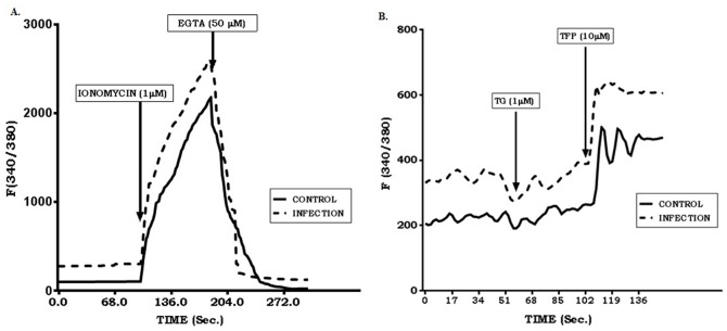
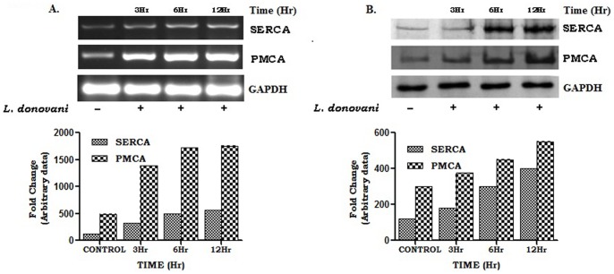
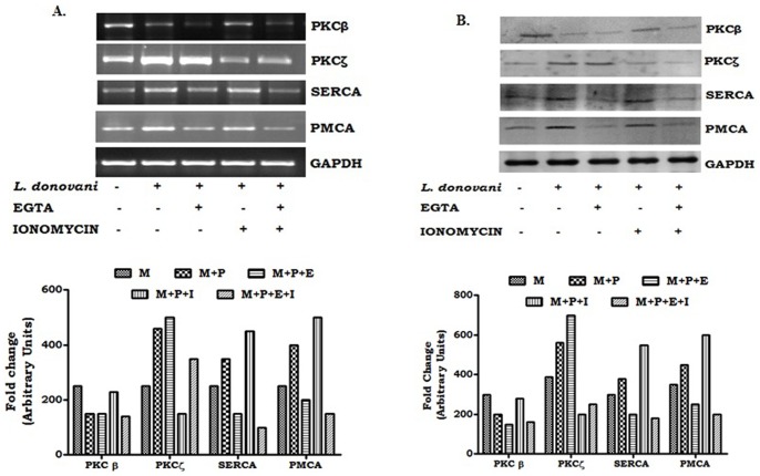
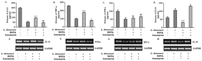
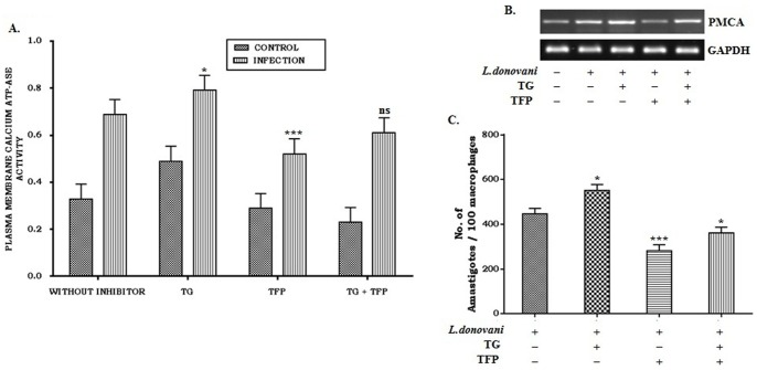
Calcium is an ubiquitous cellular signaling molecule that controls a variety of cellular processes and is strictly maintained in the cellular compartments by the coordination of various Ca2+ pumps and channels. Two such fundamental calcium pumps are plasma membrane calcium ATPase (PMCA) and Sarco/endoplasmic reticulum calcium ATPase (SERCA) which play a pivotal role in maintaining intracellular calcium homeostasis. This intracellular Ca2+ homeostasis is often disturbed by the protozoan parasite Leishmania donovani, the causative organism of visceral leishmaniasis. In the present study we have dileneated the involvement of PMCA4 and SERCA3 during leishmaniasis. We have observed that during leishmaniasis, intracellular Ca2+ concentration was up-regulated and was further controlled by both PMCA4 and SERCA3. Inhibition of these two Ca2+-ATPases resulted in decreased parasite burden within the host macrophages due to enhanced intracellular Ca2+. Contrastingly, on the other hand, activation of PMCA4 was found to enhance the parasite burden. Our findings also highlighted the importance of Ca2+ in the modulation of cytokine balance during leishmaniasis. These results thus cumulatively suggests that these two Ca2+-ATPases play prominent roles during visceral leishmaniasis.


Conflict of interest statement

Competing Interests: The authors have declared that no competing interest exists.

Figures

Figure 1
Figure 1. Detection of intracellular Ca2+ in Leishmania donovani infected macrophages in presence of different chemical agents.
Uninfected (2×10⁶ cells) and Leishmania donovani infected macrophages (1∶10) were treated with Fura-2/AM (5 µM). Geometric mean fluorescence intensity was monitored at different time intervals by flow cytometry with the addition of ionomycin (1 µM), EGTA (50 µM) (A) and thapsigargin (TG, 1 µM), trifluoperazine (TFP, 10 µM) (B). Data shown is a representative one from three different experiments conducted under identical conditions.
Figure 2
Figure 2. Expression of SERCA3 and PMCA4 at different time periods of infection.
Macrophages (2×10⁶ cells) were infected with L. donovani (1∶10) and incubated at 37°C for 3, 6 and 12 hr. The cells were collected in Trizol for RNA extraction and semi-quantitative RT-PCR was performed. PMCA4, SERCA3 and GAPDH PCR products were resolved on an agarose gel (1.5%) and quantified densitometrically using lab software as described in Methods (A). In a separate set of experiment, cell lysates were prepared from infected macrophages, followed by Western blot for PMCA4, SERCA3 and GAPDH (B). Data shown above (A and B) is a representative one experiment, performed at least three times.
Figure 3
Figure 3. Effects of EGTA and ionomycin on mRNA and protein expression of PKC-β, PKC-ζ, SERCA3 and PMCA4 in Leishmania-infected macrophages.
Macrophages (2×10⁶ cells) were infected with L. donovani (1∶10) and with 2 mM EGTA and/or 1 µM Ionomycin respectively. After 4 hr, cells were washed and incubated for another 3 hr at 37°C. The cells were collected in Trizol for RNA extraction and semi-quantitative RT-PCR was performed. PKC-β, PKC-ζ, SERCA3, PMCA4, and GAPDH PCR products were resolved on an agarose gel (1.5%) and quantified densitometrically using lab software as described in Methods (A). In a similar experiment, cell lysates were prepared from EGTA and/or ionomycin treated infected macrophages, followed by Western blot for PKC-β, PKC-ζ, SERCA3, PMCA4, and GAPDH (B). Data presented above (A and B) is a representative one of three experiments conducted under identical conditions.
Figure 4
Figure 4. Effect of EGTA and ionomycin on the expression of pro- and anti-inflammatory cytokines in L. donovani infected macrophages.
Macrophages (2×10⁶ cells) were infected with L. donovani (1∶10) and incubated at 37°C in the presence of 2 mM EGTA and/or 1 µM ionomycin. The cell supernatants were collected after 24 hr of infection. Cytokine assay was carried out by ELISA (A, B, C and D). Data represent means ± SD for 3 sets of experiments ***P<.001, *P<.05 and ns = non-significant for the comparison with infected one. In a parallel experiment, EGTA or Ionomycin treated infected cells were collected in Trizol for RNA extraction and semi-quantitative RT-PCR was performed. IL-12, TNF-α, IFN-γ, IL-10 and GAPDH PCR products were resolved on an agarose gel (1.5%) and quantified densitometrically using lab software as described in Methods. Data is from one representative experiment performed at least three times (E, F, G, and H).
Figure 5
Figure 5. Effect of inhibitors on plasma membrane Ca2+-ATPases during Leishmania infection.
Macrophages were treated with thapsigargin (TG, 1 µM) and trifluoperazine (TFP, 10 µM) followed by infection with L.donovani in 1∶10 ratio. Unbound parasites were washed off after 4 hr and further incubated for 20 hr. Cells were collected, plasma membrane was isolated and Ca2+-ATPase activity was measured. (A). In a similar experiment, inhibitor treated and infected macrophages were collected in Trizol for RNA extraction and semi-quantitative RT-PCR were performed (B). Macrophages were seeded on 8 well chamber slides (10,000 cells/well) and treated with inhibitors followed by infection with L.donovani in a 1∶10 ratio. Cells were incubated for 24 hr, fixed with chilled methanol, and stained with Giemsa. Amastigotes were then microscopically counted. ***P<.001, *P<.05 and ns = non-significant for the comparison with infected one (C). Data represented means ± SD from three different experiments (A and C). Result shown in B is a representative one from three different experiments.

Similar articles

Cited by

References

    1. Murray HW, Spitalny GL, Nathan CF (1985) Activation of mouse peritoneal macrophages in vitro and in vivo by interferon-gamma. J Immunol 134: 1619–1622. - PubMed
    1. Wilmann M, Gautel M, Mayans O (2000) Activation of calcium/calmodulin regulated kinases. Cell Mol Biol 46(5): 883–94. - PubMed
    1. Olivier M, Gregory DJ, Forget G (2005) Subversion mechanisms by which Leishmania parasites can escape the host immune response: A signaling point of view. Clin Microbiol Rev 18(2): 293–305. - PMC - PubMed
    1. Descoteaux A, Matlashewski G (1989) c-fos and tumor necrosis factor gene expression in Leishmania donovani-infected macrophages. Mol Cell Biol 9: 5223–7. - PMC - PubMed
    1. Nishizuka (1988) Studies and prospectives of protein kinase C in signal transduction. Nippon Ketsueki Gakkai Zasshi 51(8): 1321–6. - PubMed

Publication types

MeSH terms

Substances

LinkOut - more resources