. 2014 Oct 17;9(10):e110434.
doi: 10.1371/journal.pone.0110434. eCollection 2014.

Macrophage migration inhibitory factor is involved in ectopic endometrial tissue growth and peritoneal-endometrial tissue interaction in vivo: a plausible link to endometriosis development

Affiliations

Macrophage migration inhibitory factor is involved in ectopic endometrial tissue growth and peritoneal-endometrial tissue interaction in vivo: a plausible link to endometriosis development

Halima Rakhila et al. PLoS One. .

Abstract

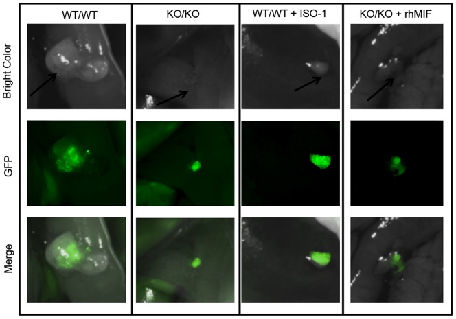
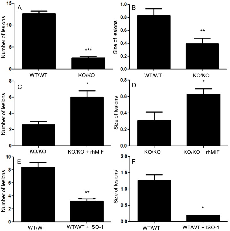
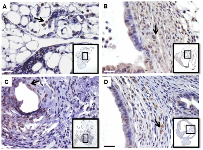
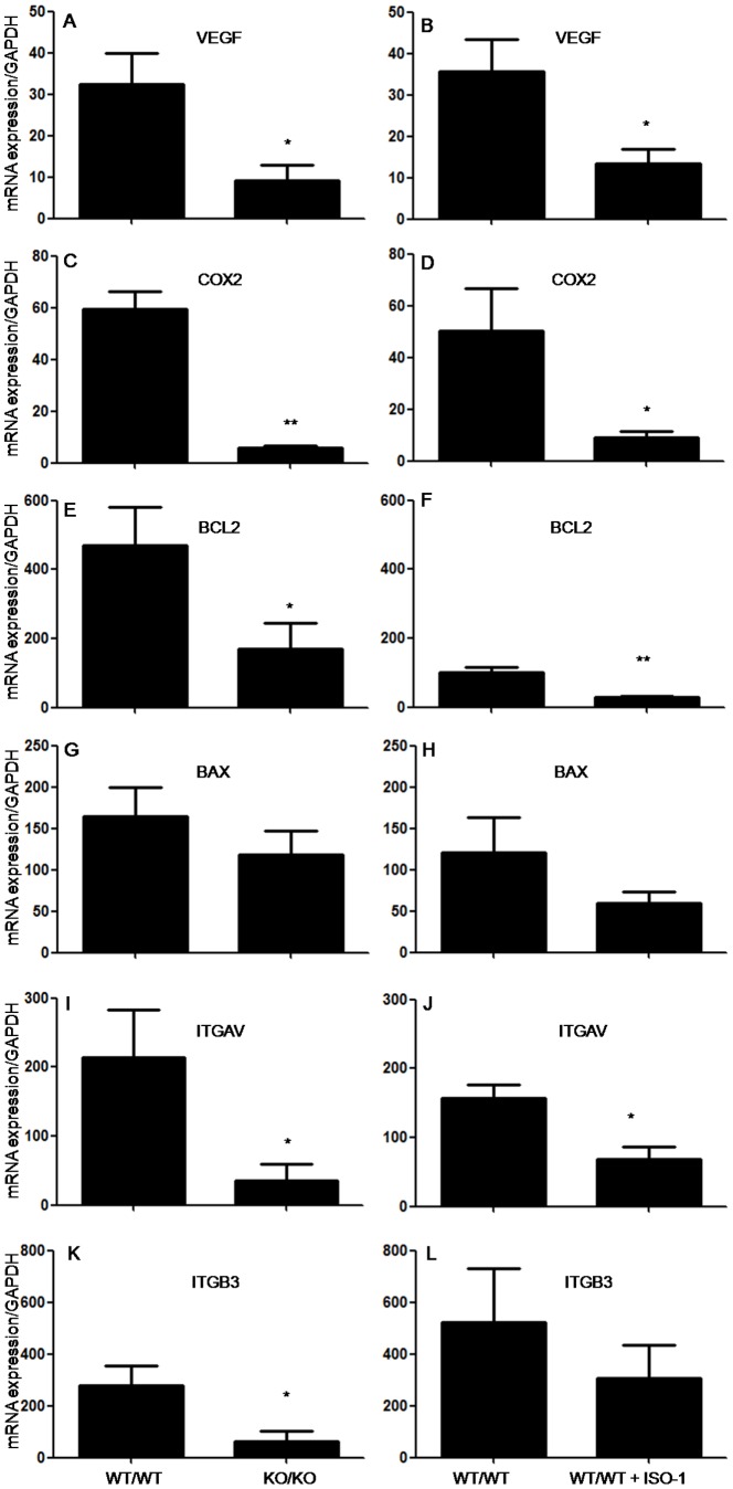
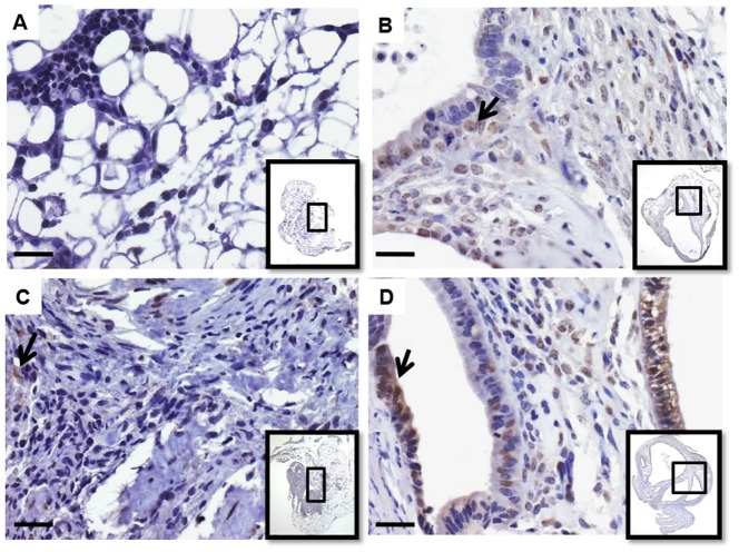
Pelvic inflammation is a hallmark of endometriosis pathogenesis and a major cause of the disease's symptoms. Abnormal immune and inflammatory changes may not only contribute to endometriosis-major symptoms, but also contribute to ectopic endometrial tissue growth and endometriosis development. A major pro-inflammatory factors found elevated in peritoneal fluid of women with endometriosis and to be overexpressed in peritoneal fluid macrophages and active, highly vascularized and early stage endometriotic lesions, macrophage migration inhibitory factor (MIF) appeared to induce angiogenic and inflammatory and estrogen producing phenotypes in endometriotic cells in vitro and to be a possible therapeutic target in vivo. Using a mouse model where MIF-knock out (KO) mice received intra-peritoneal injection of endometrial tissue from MIF-KO or syngeneic wild type (WT) mice and vice versa, our current study revealed that MIF genetic depletion resulted in a marked reduction ectopic endometrial tissue growth, a disrupted tissue structure and a significant down regulation of the expression of major inflammatory (cyclooxygenease-2), cell adhesion (αv and β3 integrins), survival (B-cell lymphoma-2) and angiogenic (vascular endothelial cell growth) factors relevant to endometriosis pathogenesis, whereas MIF add-back to MIF-KO mice significantly restored endometriosis-like lesions number and size. Interestingly, cross-experiments revealed that MIF presence in both endometrial and peritoneal host tissues is required for ectopic endometrial tissue growth and pointed to its involvement in endometrial-peritoneal interactions. This study provides compelling evidence for the role of MIF in endometriosis development and its possible interest for a targeted treatment of endometriosis.


Conflict of interest statement

Competing Interests: The authors have declared that no competing interests exist.

Figures

Figure 1
Figure 1. Stereomicroscopic observation of endometriosis-like lesions.
Endometrial implants as labeled with CFDA-SE and observed at sacrifice by fluorescence stereomicroscopy in MIF-KO mice inoculated with endometrial tissue from syngeneic MIF-KO mice (KO/KO) or WT mice inoculated with syngeneic WT murine endometrial tissue (WT/WT) (controls). Endometrial implants in KO/KO mice treated with rhMIF (0.008 mg/kg) or WT/WT mice treated with ISO-1 (4 mg/kg) are shown. Implants were observed by optic light or epi-fluorescence (GFP). Merged images are shown.
Figure 2
Figure 2. Development of endometriosis-like lesions in KO/KO mice and control WT/WT mice.
Endometriosis-like lesions in KO/KO mice and control WT/WT mice were numbered and their sizes measured under fluorescence stereomicroscopy at sacrifice (A, B). Parallel experiments were performed where rhMIF (0.008 mg/kg) was added back to KO/KO mice (C, D), while ISO-1 (4 mg/kg) was used to treat WT/WT mice (E, F). Data are from 5 or 6 mice per group and expressed as mean ± SEM;*, p< 0.05, **, p< 0.01 and ***, p< 0.001 compared with the corresponding control group with the unpaired t-test.
Figure 3
Figure 3. Effect of endometrial tissue inoculation and treatment on the body weight and survival rate of animals.
Mice were treated with ISO-1 (4 mg/kg) (A, B), rhMIF (0.008 mg/kg) (C, D). Control mice were treated with the vehicle (PBS). Data are means ± SEM from 5 mice treated with ISO-1, 5 mice treated with rhMIF and 10 mice treated with the vehicle.
Figure 4
Figure 4. Immunostaining and histological examination of murine endometrial implants.
Immunostaining of endometrial tissue from WT mice before (A, black arrows) and after inoculation and implantation into WT mice (controls) (B, black arrows) showing CK7-positive epithelial glands. ES, endometrial stroma; EG, endometrial glands;HT, host tissue. Hematoxylin-eosin staining of endometrial implants from KO/KO mice(C), WT/WT mice (D) and mice treated with ISO-1 (E) or rhMIF (F). Insets show general histological views of endometrial implants. Scale bar,10 µm.
Figure 5
Figure 5. Immunostaining of MIF receptor CD74 in murine endometrial implants.
CD74 immunostaining was carried out on implants from KO/KO mice (A), WT/WT mice (B) and mice treated with ISO-1 (C) or rhMIF (D). Insets show general histological views of endometrial implants. Black arrows show CD74 positive cells. Scale bar, 10 µm.
Figure 6
Figure 6. Real-time PCR analysis of the expression of genes mainly involved in cell adhesion and vascularisation of endometriosis-like lesions.
Histogram representation of the effect of MIF genetic depletion or antagonism versus controls on VEGF (A, B), COX2 (C, D), BCL2 (E, F), BAX (G, H), ITGAV (I, J) and ITGB3 (K, L) mRNA expression in endometriosis-like lesions by quantitative real time PCR. For each factor, the ratio of mRNA level to GAPDH mRNA was determined. Results were from WT and KOmice (n  =  6) with no treatment (controls) and from WT treated with ISO-1 (n  =  5). Data are mean ± SEM; *, p <0.05 and **, p < 0.01 as compared to the control group.
Figure 7
Figure 7. Immunostaining of PCNA in murine endometrial implants.
PCNA immunostainingwas carried out on endometrial implants from KO/KO mice (A), WT/WT mice (B) and mice treated with ISO-1 (C) or rhMIF (D). Insets show general histological views of endometrial implants. Black arrows show CD74 positive cells. Scale bar,10 µm.

Similar articles

Cited by

References

    1. Sampson J (1927) Peritoneal endometriosis due to menstrual dissemination of endometrial tisue into the peritoneal cavity. Am J Obstet Gynecol 14: 422–469.
    1. Berbic M, Fraser IS (2013) Immunology of normal and abnormal menstruation. Womens Health (Lond Engl) 9: 387–395. - PubMed
    1. Khoufache K, Michaud N, Harir N, Kibangou Bondza P, Akoum A (2012) Anomalies in the inflammatory response in endometriosis and possible consequences: a review. Minerva Endocrinol 37: 75–92. - PubMed
    1. Veillat V, Sengers V, Metz CN, Roger T, Leboeuf M, et al. (2012) Macrophage migration inhibitory factor is involved in a positive feedback loop increasing aromatase expression in endometriosis. Am J Pathol 181: 917–927. - PubMed
    1. Leng L, Bucala R (2006) Insight into the biology of macrophage migration inhibitory factor (MIF) revealed by the cloning of its cell surface receptor. Cell Res 16: 162–168. - PubMed

Publication types

MeSH terms

Substances