

Resting-state functional connectivity modulation and sustained changes after real-time functional magnetic resonance imaging neurofeedback training in depression
- PMID: 25329241
- PMCID: PMC4238245
- DOI: 10.1089/brain.2014.0262
Resting-state functional connectivity modulation and sustained changes after real-time functional magnetic resonance imaging neurofeedback training in depression
Abstract
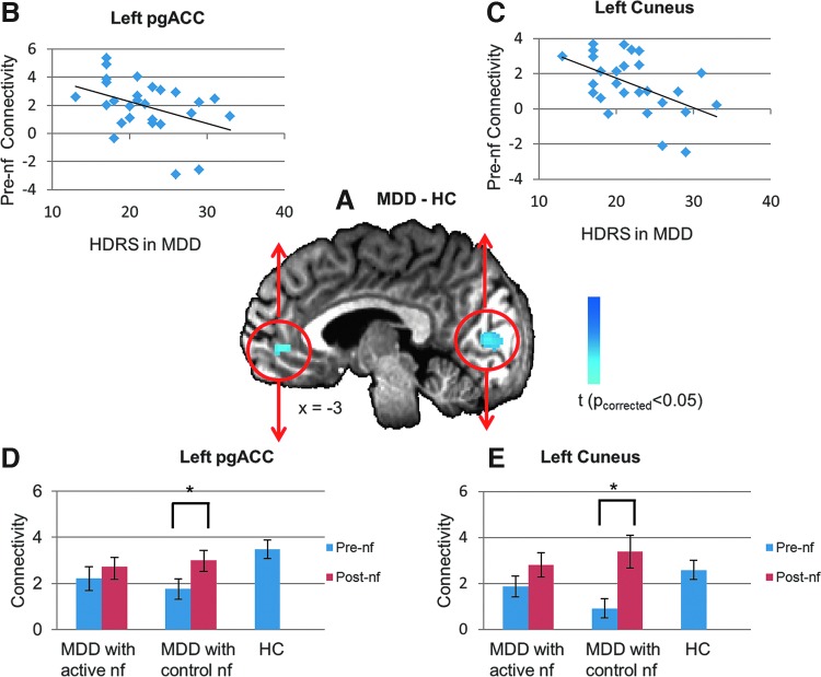
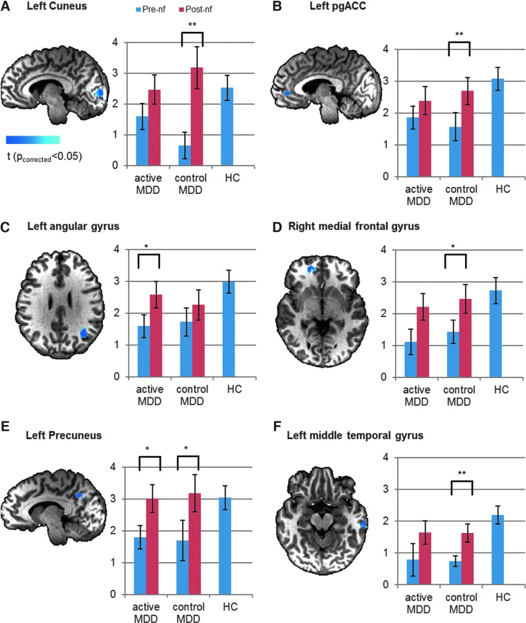
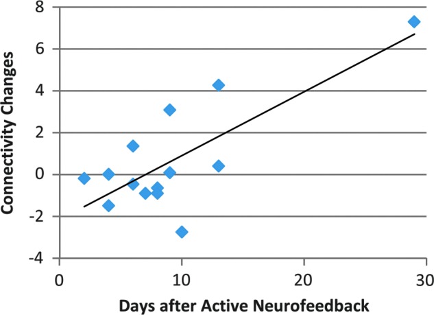
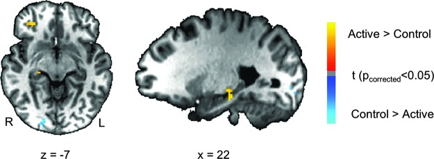
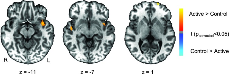
Amygdala hemodynamic responses to positive stimuli are attenuated in major depressive disorder (MDD) and normalize with remission. Real-time functional magnetic resonance imaging neurofeedback (rtfMRI-nf) training with the goal of upregulating amygdala activity during recall of happy autobiographical memories (AMs) has been suggested, and recently explored, as a novel therapeutic approach that resulted in improvement in self-reported mood in depressed subjects. In this study, we assessed the possibility of sustained brain changes as well as the neuromodulatory effects of rtfMRI-nf training of the amygdala during recall of positive AMs in MDD and matched healthy subjects. MDD and healthy subjects went through one visit of rtfMRI-nf training. Subjects were assigned to receive active neurofeedback from the left amygdale (LA) or from a control region putatively not modulated by AM recall or emotion regulation, that is, the left horizontal segment of the intraparietal sulcus. To assess lasting effects of neurofeedback in MDD, the resting-state functional connectivity before and after rtfMRI-nf in 27 depressed subjects, as well as in 27 matched healthy subjects before rtfMRI-nf was measured. Results show that abnormal hypo-connectivity with LA in MDD is reversed after rtfMRI-nf training by recalling positive AMs. Although such neuromodulatory changes are observed in both MDD groups receiving feedback from respective active and control brain regions, only in the active group are larger decreases of depression severity associated with larger increases of amygdala connectivity and a significant, positive correlation is found between the connectivity changes and the days after neurofeedback. In addition, active neurofeedback training of the amygdala enhances connectivity with temporal cortical regions, including the hippocampus. These results demonstrate lasting brain changes induced by amygdala rtfMRI-nf training and suggest the importance of reinforcement learning in rehabilitating emotion regulation in depression.
Keywords: amygdala; functional connectivity; major depressive disorder; neurofeedback; real-time fMRI; resting state.
Figures
Similar articles
-
Altered task-based and resting-state amygdala functional connectivity following real-time fMRI amygdala neurofeedback training in major depressive disorder.Neuroimage Clin. 2017 Dec 5;17:691-703. doi: 10.1016/j.nicl.2017.12.004. eCollection 2018. Neuroimage Clin. 2017. PMID: 29270356 Free PMC article.
-
Real-time FMRI neurofeedback training of amygdala activity in patients with major depressive disorder.PLoS One. 2014 Feb 11;9(2):e88785. doi: 10.1371/journal.pone.0088785. eCollection 2014. PLoS One. 2014. PMID: 24523939 Free PMC article.
-
Correlation between amygdala BOLD activity and frontal EEG asymmetry during real-time fMRI neurofeedback training in patients with depression.Neuroimage Clin. 2016 Feb 12;11:224-238. doi: 10.1016/j.nicl.2016.02.003. eCollection 2016. Neuroimage Clin. 2016. PMID: 26958462 Free PMC article.
-
Amygdala real-time functional magnetic resonance imaging neurofeedback for major depressive disorder: A review.Psychiatry Clin Neurosci. 2018 Jul;72(7):466-481. doi: 10.1111/pcn.12665. Epub 2018 May 21. Psychiatry Clin Neurosci. 2018. PMID: 29687527 Free PMC article. Review.
-
Amygdala Modulation During Emotion Regulation Training With fMRI-Based Neurofeedback.Front Hum Neurosci. 2019 Mar 26;13:89. doi: 10.3389/fnhum.2019.00089. eCollection 2019. Front Hum Neurosci. 2019. PMID: 30971906 Free PMC article.
Cited by
-
The Evolutionary Theory of Depression.Med Sci Monit. 2017 May 13;23:2267-2274. doi: 10.12659/msm.901240. Med Sci Monit. 2017. PMID: 28500855 Free PMC article. Review.
-
Characteristics of brain functional and structural connectivity in alexithymic students.Neuropsychiatr Dis Treat. 2018 Oct 9;14:2609-2615. doi: 10.2147/NDT.S174015. eCollection 2018. Neuropsychiatr Dis Treat. 2018. PMID: 30349259 Free PMC article.
-
Attentional bias in depression: understanding mechanisms to improve training and treatment.Curr Opin Psychol. 2019 Oct;29:266-273. doi: 10.1016/j.copsyc.2019.07.036. Epub 2019 Jul 31. Curr Opin Psychol. 2019. PMID: 31521030 Free PMC article. Review.
-
Alterations of amygdala-prefrontal connectivity with real-time fMRI neurofeedback in BPD patients.Soc Cogn Affect Neurosci. 2016 Jun;11(6):952-60. doi: 10.1093/scan/nsw016. Epub 2016 Feb 1. Soc Cogn Affect Neurosci. 2016. PMID: 26833918 Free PMC article.
-
Clinical response to neurofeedback in major depression relates to subtypes of whole-brain activation patterns during training.Mol Psychiatry. 2025 Jun;30(6):2707-2717. doi: 10.1038/s41380-024-02880-3. Epub 2024 Dec 26. Mol Psychiatry. 2025. PMID: 39725743 Free PMC article.
References
-
- Addis DR, McIntosh AR, Moscovitch M, Crawley AP, McAndrews MP. 2004. Characterizing spatial and temporal features of autobiographical memory retrieval networks: a partial least squares approach. Neuroimage 23:1460–1471 - PubMed
-
- Allison T, Puce A, McCarthy G. 2000. Social perception from visual cues: role of the STS region. Trends Cogn Sci 4:267–278 - PubMed
-
- American Psychological Association (APA). 2000. Diagnostic and Statistical Manual of Mental Disorders, Fourth Edition, Text Revision. Washington, DC: American Psychiatric Association
-
- Baas D, Aleman A, Kahn RS. 2004. Lateralization of amygdala activation: a systematic review of functional neuroimaging studies. Brain Res Rev 45:96–103 - PubMed
-
- Birn RM, Diamond JB, Smith MA, Bandettini PA. 2006. Separating respiratory-variation-related fluctuations from neuronal-activity-related fluctuations in fMRI. Neuroimage 31:1536–1548 - PubMed
Publication types
MeSH terms
Substances
LinkOut - more resources
Full Text Sources
Other Literature Sources
Medical
