. 2014 Oct 20;9(10):e111093.
doi: 10.1371/journal.pone.0111093. eCollection 2014.

Bestrophin 3 ameliorates TNFα-induced inflammation by inhibiting NF-κB activation in endothelial cells

Affiliations

Bestrophin 3 ameliorates TNFα-induced inflammation by inhibiting NF-κB activation in endothelial cells

Wei Song et al. PLoS One. .

Abstract

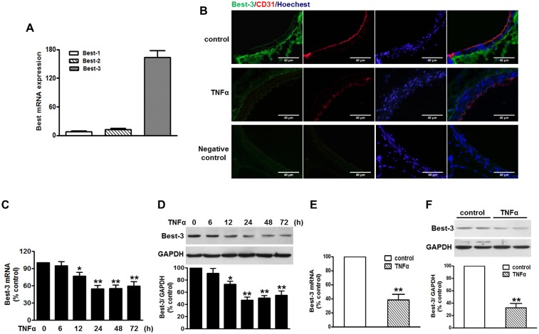
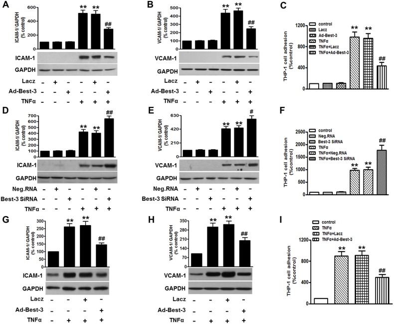
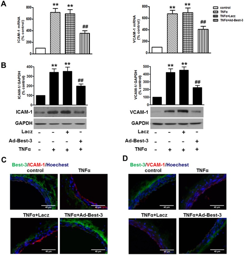
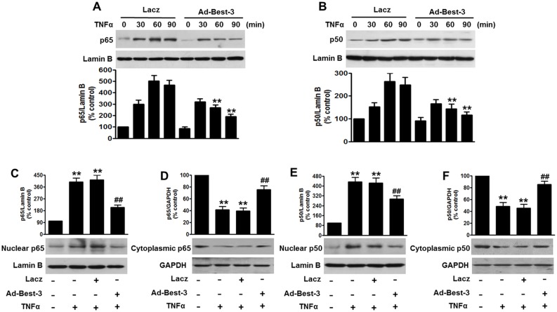
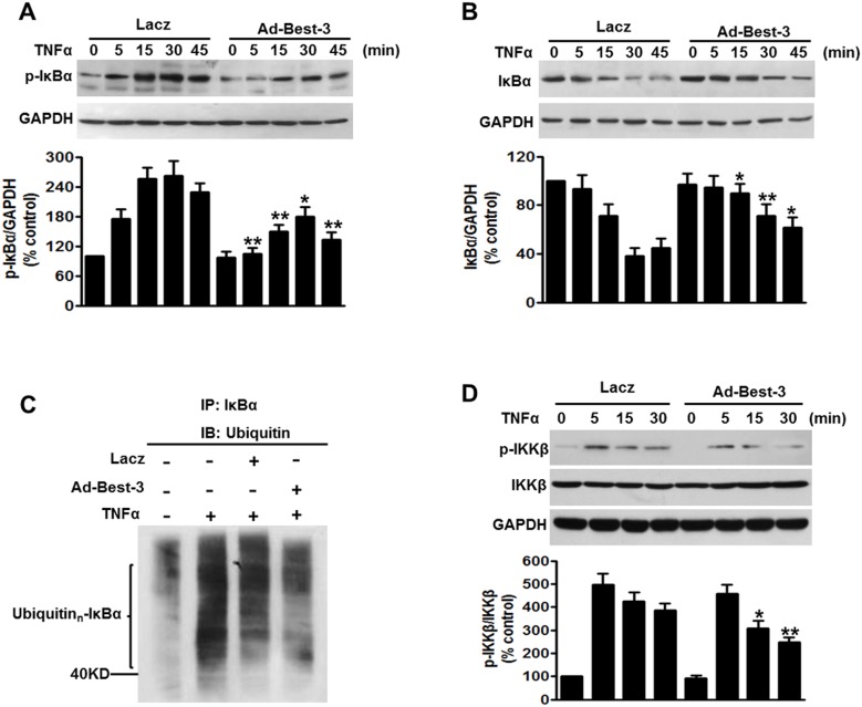
Increasing evidences have suggested vascular endothelial inflammatory processes are the initiator of atherosclerosis. Bestrophin 3 (Best-3) is involved in the regulation of cell proliferation, apoptosis and differentiation of a variety of physiological functions, but its function in cardiovascular system remains unclear. In this study, we investigated the effect of Best-3 on endothelial inflammation. We first demonstrated that Best-3 is expressed in endothelial cells and decreased after tumor necrosis factor-α (TNFα) challenge. Overexpression of Best-3 significantly attenuated TNFα-induced expression of adhesion molecules and chemokines, and subsequently inhibited the adhesion of monocytes to human umbilical vein endothelial cells (HUVECs). Conversely, knockdown of Best-3 with siRNA resulted in an enhancement on TNFα-induced expression of adhesion molecules and chemokines and adhesion of monocytes to HUVECs. Furthermore, overexpression of Best-3 with adenovirus dramatically ameliorated inflammatory response in TNFα-injected mice. Mechanistically, we found up-regulation of Best-3 inhibited TNFα-induced IKKβ and IκBα phosphorylation, IκBα degradation and NF-κB translocation. Our results demonstrated that Best-3 is an endogenous inhibitor of NF-κB signaling pathway in endothelial cells, suggesting that forced Best-3 expression may be a novel approach for the treatment of vascular inflammatory diseases.


Conflict of interest statement

Competing Interests: The authors have declared that no competing interests exist.

Figures

Figure 1
Figure 1. TNFα decreased Best-3 expression in endothelium.
(A) quantitative PCR analysis of Best-1, Best-2 and Best-3 mRNA expression in HUVECs. n = 6. (B) immunofluorescent staining of Best-3 (green) and CD31 (red) and Hoechst (blue) in thoracic aorta of mice after TNFα challenge for 3 days. n = 4–6. Scale bars, 40 µm. (C, D) HUVECs were stimulated with TNFα (10 ng/ml) for different times as indicated. Quantitative PCR (C) and western blot (D) results show a time-dependent decrease of Best-3 expression. *P<0.05, **P<0.01 vs. untreated group, n = 5. (E, F) quantitative PCR (E) and western blot (F) analysis of Best-3 expression in MAECs isolated from mice after treatment mentioned in method section, respectively. **P<0.01 vs. control group, n = 8.
Figure 2
Figure 2. Best-3 ameliorated TNFα-induced inflammatory response in endothelial cells.
(A, B) HUVECs were transfected with Lacz or Ad-Best-3 for 48 h prior to TNFα treatment for 24 h. ICAM-1 (A) and VCAM-1 (B) were examined by western blot, respectively. (C) after treatment mentioned in (A, B), adhesion of VibrantDiO-labeled THP-1 to HUVECs were analyzed. (D, E) HUVECs were transfected with negative siRNA (Neg. RNA) or Best-3 siRNA for 48 h prior to TNFα incubation. ICAM-1 (D) and VCAM-1 (E) were detected by western blot, respectively. (F) after treatment mentioned in (D, E), adhesion of THP-1 to HUVECs was analyzed. (G, H) western blot detection of ICAM-1 (G) and VCAM-1 (H) expressions in MAECs isolated form mice after treatment mentioned in method section. (I) adhesion of THP-1 to MAECs was analyzed. All data are presented as mean ± SEM. **P<0.01 vs. control, #P<0.05, ##P<0.01 vs. TNFα alone, n = 6.
Figure 3
Figure 3. Reduced inflammatory response in Ad-Best-3-infected mice.
C57BL/6 mice were infected with Ad-Best-3 (109 pfu/mouse) or Lacz (109 pfu/mouse) for 1 week in the presence of TNFα (30 µg/kg) for another 3 days. (A. B) the expression of ICAM-1 and VCAM-1 in the aorta was analyzed by quantitative PCR (A, upper panel) and western blot (B, middle panel), respectively. **P<0.01 vs. control, ##P<0.01 vs. TNFα alone, n = 7. (C, D) immunofluorescent staining of Best-3 (green) and ICAM-1 (red) and Hoechst (blue) (C), or Best-3 (green) and VCAM-1 (red) and Hoechst (blue) (D) in thoracic aorta of mice, respectively. Scale bars, 40 µm. n = 4–6.
Figure 4
Figure 4. Best-3 repressed TNFα-induced NF-κB activation in endothelial cells.
(A, B) HUVECs were infected with Lacz or Ad-Best-3 for 48 h, and then incubated with TNFα (10 ng/ml) for different times as indicated. Nuclear fractions were isolated and detected by western blot using p65 (A) and p50 (B) antibodies. **P<0.01 vs. similarity treated control, n = 4. (C–F) nuclear and cytoplasmic fractions of MAECs isolated from mice after treatment mentioned in method section were analyzed by western blot to detect the expressions of p65 (C, D) and p50 (E, F). **P<0.01 vs. control, ##P<0.01 vs. TNFα alone, n = 6.
Figure 5
Figure 5. Best-3 suppressed IKKβ/IκBα pathway to inhibit inflammation.
(A, B) HUVECs were infected with Lacz or Ad-Best-3 for 48 h, and then incubated with TNFα (10 ng/ml) for different times as indicated. Cell lysates were subjected to western blot analysis using p-IκBα (A) and IκBα (B) antibodies. *P<0.05, **P<0.01 vs. similarity treated control, n = 4. (C) western blot analysis of ubiquitinated IκBα in HUVECs treated with Lazc or Ad-Best-3 in the presence of TNFα for 30 min. Cell lysates were immunoprecipitated with IκBα antibody and immunoprecipitated proteins were blotted with ubiquitin antibody to reveal ubiquitination of IκBα. (D) western blot analysis of p-IKKβ and IKKβ of HUVECs treated with Lacz or Ad-Best-3 for 48 h in the presence of TNFα for different times as indicated. *P<0.05, **P<0.01 vs. similarity treated control, n = 4.

References

    1. Ross R (1999) Atherosclerosis is an inflammatory disease. Am Heart J 138: S419–420. - PubMed
    1. Schiffrin EL (2002) Canadian Institutes of Health Research Multidisciplinary Research Group on Hypertension (2002) Beyond blood pressure: the endothelium and atherosclerosis progression. Am J Hypertens 15: 115S–122S. - PubMed
    1. van den Oever IA, Raterman HG, Nurmohamed MT, Simsek S (2010) Endothelial dysfunction, inflammation, and apoptosis in diabetes mellitus. Mediators Inflamm 2010: 792393. - PMC - PubMed
    1. Davignon J, Ganz P (2004) Role of endothelial dysfunction in atherosclerosis. Circulation 109: III27–III32. - PubMed
    1. Sun X, Icli B, Wara AK, Belkin N, He S, et al. (2012) MicroRNA-181b regulates NF-κB-mediated vascular inflammation. J Clin Invest 122: 1973–1990. - PMC - PubMed

Publication types

MeSH terms

LinkOut - more resources