

Contrasting roles for MyoD in organizing myogenic promoter structures during embryonic skeletal muscle development
- PMID: 25329411
- PMCID: PMC4276533
- DOI: 10.1002/dvdy.24217
Contrasting roles for MyoD in organizing myogenic promoter structures during embryonic skeletal muscle development
Abstract
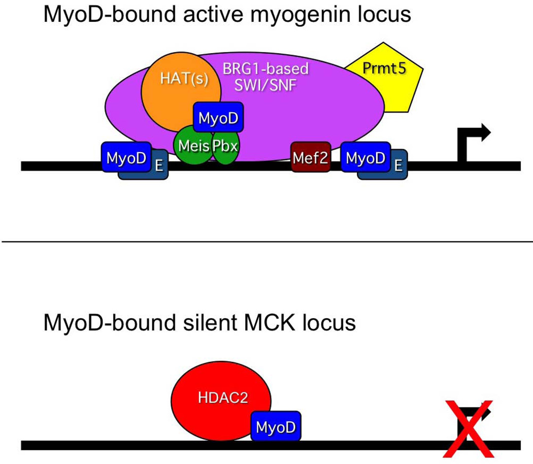
Background: Among the complexities of skeletal muscle differentiation is a temporal distinction in the onset of expression of different lineage-specific genes. The lineage-determining factor MyoD is bound to myogenic genes at the onset of differentiation whether gene activation is immediate or delayed. How temporal regulation of differentiation-specific genes is established remains unclear.
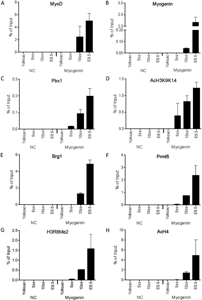
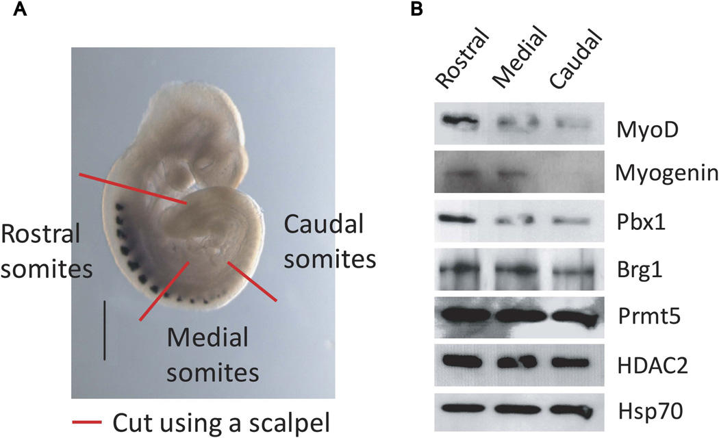
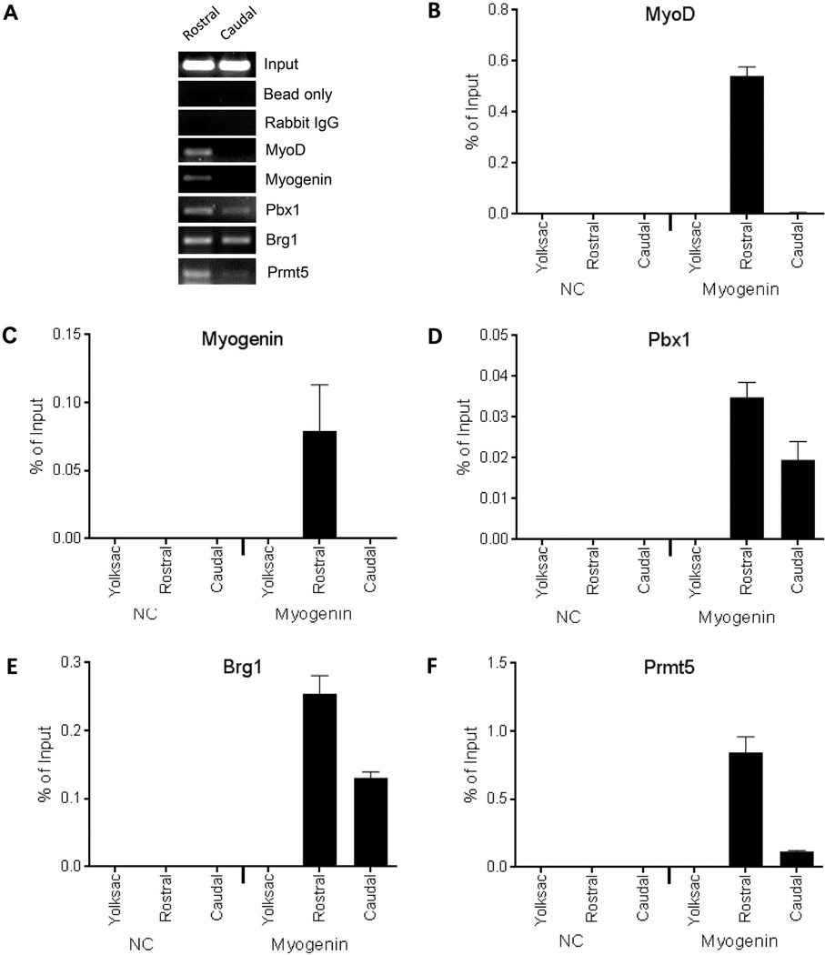
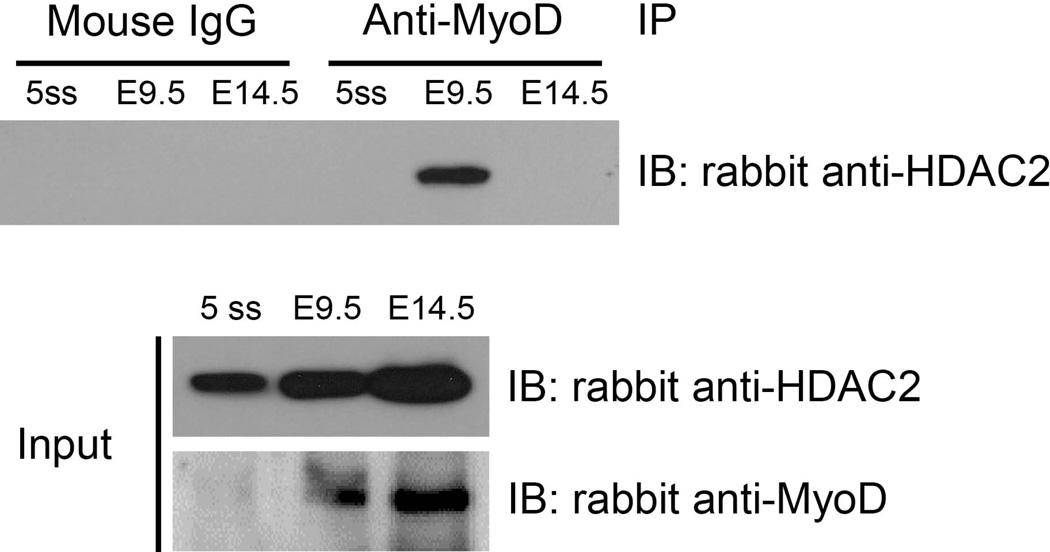
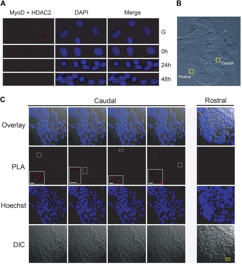
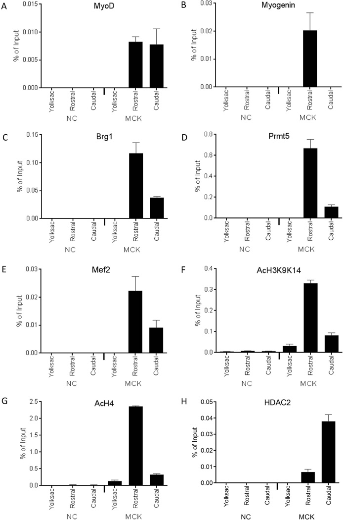
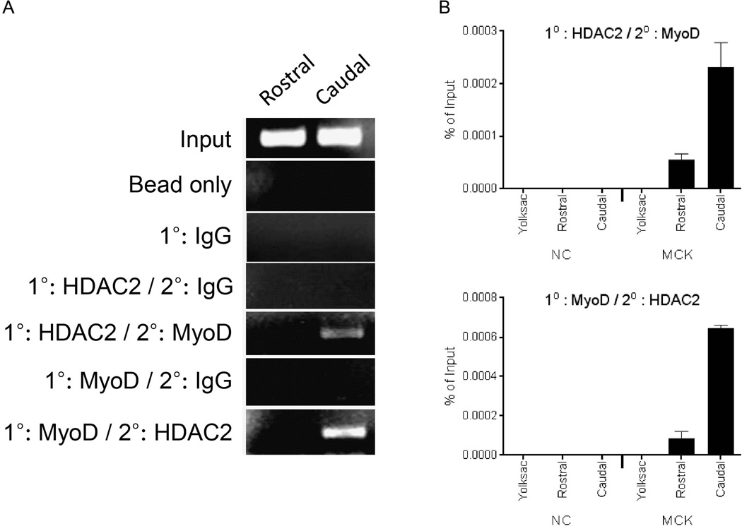
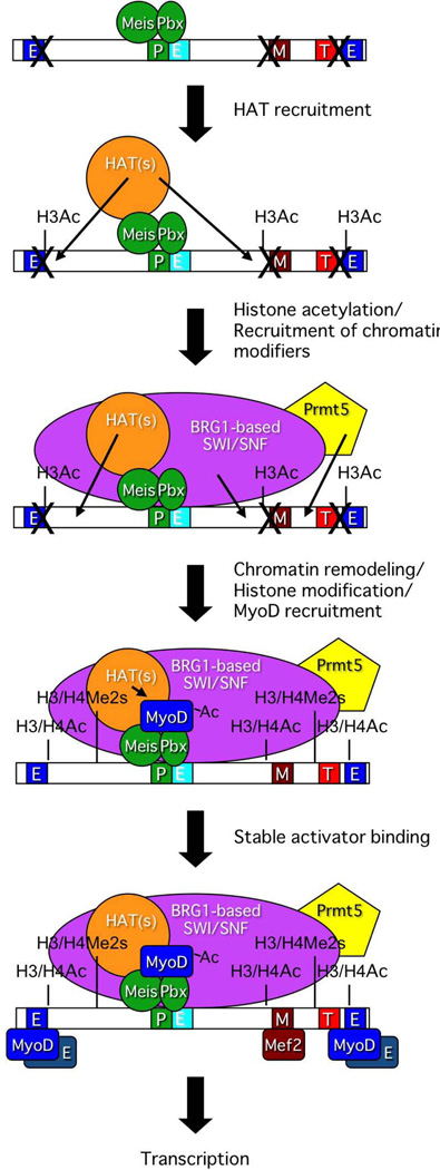
Results: Using embryonic tissue, we addressed the molecular differences in the organization of the myogenin and muscle creatine kinase (MCK) gene promoters by examining regulatory factor binding as a function of both time and spatial organization during somitogenesis. At the myogenin promoter, binding of the homeodomain factor Pbx1 coincided with H3 hyperacetylation and was followed by binding of co-activators that modulate chromatin structure. MyoD and myogenin binding occurred subsequently, demonstrating that Pbx1 facilitates chromatin remodeling and modification before myogenic regulatory factor binding. At the same time, the MCK promoter was bound by HDAC2 and MyoD, and activating histone marks were largely absent. The association of HDAC2 and MyoD was confirmed by co-immunoprecipitation, proximity ligation assay (PLA), and sequential ChIP.
Conclusions: MyoD differentially promotes activated and repressed chromatin structures at myogenic genes early after the onset of skeletal muscle differentiation in the developing mouse embryo.
Keywords: Pbx; gene expression; gene regulation; histone deacetylase; myogenesis; somite.
© 2014 Wiley Periodicals, Inc.
Figures
Similar articles
-
Pbx marks genes for activation by MyoD indicating a role for a homeodomain protein in establishing myogenic potential.Mol Cell. 2004 May 21;14(4):465-77. doi: 10.1016/s1097-2765(04)00260-6. Mol Cell. 2004. PMID: 15149596
-
Skeletal muscle specification by myogenin and Mef2D via the SWI/SNF ATPase Brg1.EMBO J. 2006 Feb 8;25(3):490-501. doi: 10.1038/sj.emboj.7600943. Epub 2006 Jan 19. EMBO J. 2006. PMID: 16424906 Free PMC article.
-
MyoD targets chromatin remodeling complexes to the myogenin locus prior to forming a stable DNA-bound complex.Mol Cell Biol. 2005 May;25(10):3997-4009. doi: 10.1128/MCB.25.10.3997-4009.2005. Mol Cell Biol. 2005. PMID: 15870273 Free PMC article.
-
Myogenic regulatory factors and the specification of muscle progenitors in vertebrate embryos.Annu Rev Cell Dev Biol. 2002;18:747-83. doi: 10.1146/annurev.cellbio.18.012502.105758. Epub 2002 Apr 2. Annu Rev Cell Dev Biol. 2002. PMID: 12142270 Review.
-
Function of the myogenic regulatory factors Myf5, MyoD, Myogenin and MRF4 in skeletal muscle, satellite cells and regenerative myogenesis.Semin Cell Dev Biol. 2017 Dec;72:19-32. doi: 10.1016/j.semcdb.2017.11.011. Epub 2017 Nov 15. Semin Cell Dev Biol. 2017. PMID: 29127046 Review.
Cited by
-
Spatial re-organization of myogenic regulatory sequences temporally controls gene expression.Nucleic Acids Res. 2015 Feb 27;43(4):2008-21. doi: 10.1093/nar/gkv046. Epub 2015 Feb 4. Nucleic Acids Res. 2015. PMID: 25653159 Free PMC article.
-
Role of Histone Deacetylases in Skeletal Muscle Physiology and Systemic Energy Homeostasis: Implications for Metabolic Diseases and Therapy.Front Physiol. 2020 Aug 11;11:949. doi: 10.3389/fphys.2020.00949. eCollection 2020. Front Physiol. 2020. PMID: 32848876 Free PMC article. Review.
-
Pbx4 is Required for the Temporal Onset of Zebrafish Myocardial Differentiation.J Dev Biol. 2015;3(4):93-111. doi: 10.3390/jdb3040093. J Dev Biol. 2015. PMID: 26770887 Free PMC article.
-
Cysteine Rich Intestinal Protein 2 is a copper-responsive regulator of skeletal muscle differentiation and metal homeostasis.PLoS Genet. 2024 Dec 5;20(12):e1011495. doi: 10.1371/journal.pgen.1011495. eCollection 2024 Dec. PLoS Genet. 2024. PMID: 39637238 Free PMC article.
-
MyoD phosphorylation on multiple C terminal sites regulates myogenic conversion activity.Biochem Biophys Res Commun. 2016 Dec 2;481(1-2):97-103. doi: 10.1016/j.bbrc.2016.11.009. Epub 2016 Nov 4. Biochem Biophys Res Commun. 2016. PMID: 27823936 Free PMC article.
References
-
- Bergstrom DA, Penn BH, Strand A, Perry RL, Rudnicki MA, Tapscott SJ. Promoter-specific regulation of MyoD binding and signal transduction cooperate to pattern gene expression. Mol Cell. 2002;9:587–600. - PubMed
-
- Berkes CA, Bergstrom DA, Penn BH, Seaver KJ, Knoepfler PS, Tapscott SJ. Pbx marks genes for activation by MyoD indicating a role for a homeodomain protein in establishing myogenic potential. Mol Cell. 2004;14:465–477. - PubMed
-
- Berkes CA, Tapscott SJ. MyoD and the transcriptional control of myogenesis. Semin Cell Dev Biol. 2005;16:585–595. - PubMed
Publication types
MeSH terms
Substances
Grants and funding
LinkOut - more resources
Full Text Sources
Other Literature Sources
Molecular Biology Databases
