. 2015 Apr;17(4):559-78.
doi: 10.1111/cmi.12383. Epub 2014 Nov 22.

Lipid kinases are essential for apicoplast homeostasis in Toxoplasma gondii

Affiliations

Lipid kinases are essential for apicoplast homeostasis in Toxoplasma gondii

Wassim Daher et al. Cell Microbiol. 2015 Apr.

Abstract

Phosphoinositides regulate numerous cellular processes by recruiting cytosolic effector proteins and acting as membrane signalling entities. The cellular metabolism and localization of phosphoinositides are tightly regulated by distinct lipid kinases and phosphatases. Here, we identify and characterize a unique phosphatidylinositol 3 kinase (PI3K) in Toxoplasma gondii, a protozoan parasite belonging to the phylum Apicomplexa. Conditional depletion of this enzyme and subsequently of its product, PI(3)P, drastically alters the morphology and inheritance of the apicoplast, an endosymbiotic organelle of algal origin that is a unique feature of many Apicomplexa. We searched the T. gondii genome for PI(3)P-binding proteins and identified in total six PX and FYVE domain-containing proteins including a PIKfyve lipid kinase, which phosphorylates PI(3)P into PI(3,5)P2 . Although depletion of putative PI(3)P-binding proteins shows that they are not essential for parasite growth and apicoplast biology, conditional disruption of PIKfyve induces enlarged apicoplasts, as observed upon loss of PI(3)P. A similar defect of apicoplast homeostasis was also observed by knocking down the PIKfyve regulatory protein ArPIKfyve, suggesting that in T. gondii, PI(3)P-related function for the apicoplast might mainly be to serve as a precursor for the synthesis of PI(3,5)P2 . Accordingly, PI3K is conserved in all apicomplexan parasites whereas PIKfyve and ArPIKfyve are absent in Cryptosporidium species that lack an apicoplast, supporting a direct role of PI(3,5)P2 in apicoplast homeostasis. This study enriches the already diverse functions attributed to PI(3,5)P2 in eukaryotic cells and highlights these parasite lipid kinases as potential drug targets.


Figures

Figure 1
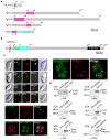
Figure 1. Expression and localization of TgPI3-Kinase in tachyzoites
(A) Depicted are the two pathways implicated in PI(3,5)P2 biosynthesis and turnover. The PI3Kinase converts PI to PI(3)P and the PAS complex (PIKfyve, ArPIKfyve and Sac3) converts PI(3)P to PI(3,5)P2. ArPIKfyve binds to the Sac3 phosphatase that reverts PI(3,5)P2 back to PI(3)P. The FYVE and PX domains containing proteins are considered as PI(3)P-recognizing effectors. (B) Schematic representation of T. gondii PI3K predicted using SMART EMBL showing the C2 (a calcium dependent membrane-targeting module that, in others proteins, binds a wide variety of molecules), the PIK domain (phosphoinositide 3-kinase family accessory domain) that has been suggested to be involved in substrate presentation, and finally the 3-kinase domain responsible for the kinase activity. Scale bar represents 300 aa. (C) Insertion of a Myc tag at the N-terminus of TgPI3K and promoter exchange by single homologous recombination at the 5′ of the gene (knock-in in RH-ku80ko strain). gNt: genomic N-terminal. (D) Western blot analysis performed on transgenic or RH-ku80ko parasite lysates probed with anti-Myc antibodies. Myc-TgPI3K is found at the expected molecular mass (314 kDa). (E) IFA performed on intracellular transgenic parasites using anti-Myc or anti-ATRX1 (apicoplast) antibodies. Der1-GFP protein was used as ER marker. Scale bars represent 2 μm.
Figure 2
Figure 2. TgPI3-Kinase conditional knock-in by promoter exchange strategy
(A) Schematic representation of the strategy used to replace the endogenous promoter of TgPI3K with the tetracycline-inducible promoter. The 5′TgPI3KUTR-DHFR-tetO7-SAG4-HA(2)NtTgPI3K plasmid contains the TgPI3K 5′ UTR (in black), the dihydrofolate reductase (DHFR) cassette (in blue) and the N-terminal genomic coding sequence of TgPI3K gene (grey) fused to two HA tags (red) under the control of the inducible tetO7SAG4 promoter (orange arrow). Black arrows represent the primers used for PCR analysis and the length of the PCR fragments is indicated. (B) PCR analysis performed on pi3ki confirming double homologous recombination. Genomic DNA from TATi1-ku80ko parasites was used as negative control. (C) Schematic representation of the crossover event that will result in the replacement of TgPI3K tetracycline inducible locus by the PI3-Kinase gene present in the recombineering modified ToxP331 cosmid in fusion with a 3 HA epitope at the 3′ end of the TgPI3K gene. CAT: chloramphenicol-acetyl-transferase. (D) Semi-quantitative RT-PCR analysis of TgPI3K expression in the wild-type, mutant and complemented parasites, preceded or not by three days of induction by ATc to regulate expression. Primers specific of the gene coding for FYVE1 were used as a loading control. (E) DD-FYVE(2)-GFP expressing pi3ki parasites were processed for IFA ± ATc and in the presence of Shield-1. The DD-FYVE(2)-GFP protein shows a punctuate staining in pi3ki parasites non-treated with ATc. A cytosolic labelling was observed when the pi3ki parasites were cultured in the presence of ATc. (F) Plaque assay of HFF monolayer infected with TATi1-ku80ko or pi3ki or pi3kiC parasites pre-treated first during 48 h with ATc. After 7 days ± ATc, the HFF were stained with Giemsa.
Figure 3
Figure 3. Phenotypic consequences of TgPI3-Kinase knock-down in pi3ki strain
(A) IFA of a representative pi3ki vacuole non-treated with ATc compared with pi3ki ATc-treated vacuoles after 4 and 5 days. Anti-ATRX1 (in green) and anti-HSP60 (in red) antibodies were used to detect the apicoplast. (B) PI3-Kinase depleted parasites showed an enlarged apicoplast. Thin section electron micrographs were taken from pi3ki that had grown for 4 days in the presence or absence of ATc. A, apicoplast; M, mitochondrion; Mc, micronemes; R, rhoptries; G, Golgi apparatus; N, nucleus; D.C, daughter cell. Scale bar, 1 μm. (C) Quantification of vacuoles showing a punctuate (P) or cytosolic (C) staining upon treatment with ATc for 1 to 5 days by visualizing the DD-FYVE(2)-GFP fluorescence. Values are means ± SD for three independent experiments. (D) Quantification of TATi1-ku80ko or pi3ki parasites harbouring either a normal or an enlarged apicoplast upon treatment with ATc for 3 to 6 days using anti-ATRX1 antibodies. (E) Quantification of TATi1-ku80ko or pi3ki parasites harbouring an apicoplast upon treatment with ATc for 3 to 7 days using anti-ATRX1 and anti-HSP60 antibodies. (F) Intracellular growth of TATi1-ku80ko, pi3ki and pi3kiC parasites cultivated in presence or absence of ATc for 6 days and allowed to invade new HFF cells. Numbers of parasites per vacuole (x-axis) were counted 24 h after inoculation. The percentages of vacuoles containing varying numbers of parasites are represented on the y- axis. (D, E and F) Values are means ± SD for three independent experiments. (G) Schematic diagram showing in the course of time the different steps leading to the delayed death phenotype of pi3ki parasites.
Figure 4
Figure 4. Repertoire of proteins containing FYVE and PX domains, their expression and their localization in T. gondii
(A and B) Schematic representation of the primary structure of the proteins highlighting their different domains; the domains have been searched with SMART (http://smart.embl-heidelberg.de). The FYVE domain is highlighted in red and the PX domain in pink. Only TgPIKfyve and TgPX4 possess additional recognizable domains. TgPIKfyve harbors a chaperonin domain (CPN) (green) known to be engaged in regulatory interactions, a CHK domain (Cys, His and Lys) (blue) and the 5-kinase catalytic domain (black) at the C-terminus responsible for the lipid kinase activity. PX4 contains 5 WD40 (blue) motifs known to coordinate multi-protein complex assemblies and binding to PI(3,5)P2 (Proikas-Cezanne et al., 2007). Scale bar represents 200 a.a or 400 a.a respectively. (C) Subcellular localization of the FYVE and PX proteins. The tagged proteins were detected using anti-HA, anti-Myc or anti-Ty antibodies. Anti-CPL and anti-ATRX1 antibodies were used to stain the vacuole compartment and the apicoplast, respectively; the GRASP-RFP and Der1-GFP proteins were used as a cis-Golgi and ER markers, respectively. Scale bars represent 2 μm. (D) Western blot analysis of transgenic or RH-ku80ko parasite lysates probed with anti-HA, anti-Myc or anti-Ty antibodies. All proteins were detected by western blotting at their expected molecular weight; 58 kDa for TgFYVE1, 205 kDa for TgPX1, 276 kDa for TgPX2, 91 kDa for TgPX3, 70 kDa for TgPX4 and 612 kDa for TgPIKfyve.
Figure 5
Figure 5. TgFYVE1 and all TgPX proteins are not critical for tachyzoite survival
(A) Plaque assay stained with Giemsa 7 days after invasion of the host cells with RH-ku80ko, fyve1-ko, px1-ki, px2-ki, px3-ko and px4-ko strains. (B) Intracellular growth assay performed by counting the numbers of parasites per vacuole (x-axis) 24 h after invasion of the host cells. The percentages of vacuoles containing varying numbers of parasites are represented on the y-axis. Values are means ± SD for three independent experiments.
Figure 6
Figure 6. TgPIKfyve knock-down by promoter replacement strategy
(A) The endogenous promoter of TgPIKfyve was replaced with the tetracycline-inducible promoter. The DHFR-tetO7-SAG4-NtTgPIKfyve plasmid contains the DHFR resistance cassette (in blue) and the N-terminal genomic coding sequence of TgPIKfyve gene (in grey, 1833 bp) under the control of the inducible tetO7SAG4 promoter (yellow arrow). The primers used for PCR analysis are indicated by black arrows and the length of the PCR fragments is specified. (B) PCR analysis performed on pikfyvei, showing that single homologous recombination had occurred. Genomic DNA from TATi1-ku80ko parasites was used as negative control. (C) Semi-quantitative RT-PCR analysis of TgPIKfyve expression in the wild-type and mutant parasites, preceded or not by two days of ATc-treatment to regulate expression. The FYVE1 gene was used as a loading control. (D) Plaque assays performed in the absence (−) or presence (+) of ATc. Parasite line name is indicated to the left.
Figure 7
Figure 7. Phenotypic consequences of TgPIKfyve knock-down in pikfyvei strain
(A) Shown are representative vacuoles of the pikfyvei strain treated for 3 and 4 days with ATc and the untreated control. Anti-ATRX1 (in green) and anti-HSP60 (in red) antibodies were used to detect the apicoplast. (B) pikfyvei parasites showed a very enlarged apicoplast in dividing and non-dividing parasites. Thin section electron micrographs were taken from pikfyvei that had grown for 3 days in the presence of ATc. A, apicoplast; Mc, micronemes; R, rhoptries; G, Golgi apparatus; N, nucleus; D.C, daughter cell; D.G., dense granule. Scale bars represent 2 μm and 500 nm respectively. (C) Quantification of TATi1-ku80ko or pikfyvei parasites harbouring an apicoplast upon treatment with ATc at days 2 to 4. (D) Intracellular growth of TATi1-ku80ko and pikfyvei cultivated in presence or absence of ATc for 4 days and allowed to invade new HFF cells. The percentage of vacuoles containing varying numbers of parasites was determined 24 h after inoculation. (C and D) Values are means ± SD for three independent experiments. (E) Schematic diagram summarizing the observed phenotype and parasite survival of the pikfyvei parasites.
Figure 8
Figure 8. The associated regulator of PIKfyve (ArPIKfyve) interferes with apicoplast morphology
(A) Model for the well established molecular organization of the PAS (PIKfyve-ArPIKfyve-Sac3) complex and its role in PI(3)P-to-PI(3,5)P2 synthesis as described in the literature. The phosphatase Sac3 and the PIKfyve kinase bind to the N-terminal and C- terminal parts of the homodimeric ArPIKfyve respectively to produce PI(3,5)P2. (B) TgArPIKfyve contains 14 HEAT repeats which are highlighted in red. The 14 HEAT repeats were predicted with the repeat finding program REP (http://www.embl-heidelberg.de/~andrade/papers/rep/search.html). (C) TgArPIKfyve knock-down by promoter replacement strategy. The endogenous promoter of TgArPIKfyve was replaced with the tetracycline-inducible promoter. The DHFR-tetO7-SAG4-NtTgArPIKfyve plasmid contains the DHFR resistance cassette (in blue) and the N-terminal genomic coding sequence of TgArPIKfyve gene (in grey, 1115 bp) under the control of the inducible tetO7SAG4 promoter (yellow arrow). The primers used for PCR analysis are indicated by black arrows and the length of the PCR fragments is specified. (D) PCR analysis performed on arpikfyvei, showing that single homologous recombination had occurred. Genomic DNA of TATi1-ku80ko parasites was used as negative control. (E) Growth analysis of either TATi1-ku80ko or arpikfyvei parasites inoculated on HFF cells and cultured for 7 days ± ATc. (F) Immunofluorescence analysis of intracellular arpikfyvei parasites non-treated or cultured for 4 days with ATc and probed with anti-HSP60 (in red) or anti-ATRX1 (in green) antibodies. The arpikfyvei parasites treated with ATc show an effect on the apicoplast shape. The apicoplast was found enlarged in the residual body of the vacuole or within the parasites. Scale bars represent 2 μm. (G) Replication assay of indicated parasites grown for 5 days in presence, or absence of ATc prior to fixation. The number of parasites per PV was determined. The knockdown of ArPIKfyve protein affected parasite replication. Values are means ± SD for three independent experiments.
Figure 9
Figure 9. Depletion of PI3-Kinase or PIKfyve did not result in obvious apicoplast protein import defects
(A and B, upper panels) Western blot analysis following the maturation of either ACP-YFP or PPP1-3HA proteins, in the pi3ki or pikfyvei mutant parasites, respectively. Parasites were grown in ATc for the times indicated. Anti-GFP or anti-HA antibodies were used to detect ACP-YFP or PPP1-3HA proteins. Note the pronounced loss of the mature forms of the proteins at days 5 and 3, respectively. The tubulin protein was used as a loading control. (A and B, lower panels) pi3ki or pikfyvei parasites were grown with (d1 to d7) or without ATc (d0) and apicoplasts were counted in 300 vacuoles for each condition. Only the mean values were plotted on the two graphs. Y-axis shows the percent of parasites that show a clearly identifiable apicoplast.

Similar articles

Cited by

References

    1. Achbarou A, Mercereau-Puijalon O, Autheman JM, Fortier B, Camus D, Dubremetz JF. Characterization of microneme proteins of Toxoplasma gondii. Mol Biochem Parasitol. 1991;47:223–233. - PubMed
    1. Agrawal S, Chung DW, Ponts N, van Dooren GG, Prudhomme J, Brooks CF, et al. An apicoplast localized ubiquitylation system is required for the import of nuclear-encoded plastid proteins. PLoS Pathog. 2013;9:e1003426. - PMC - PubMed
    1. Agrawal S, van Dooren GG, Beatty WL, Striepen B. Genetic evidence that an endosymbiont-derived endoplasmic reticulum-associated protein degradation (ERAD) system functions in import of apicoplast proteins. J Biol Chem. 2009;284:33683–33691. - PMC - PubMed
    1. Balla T. Phosphoinositides: tiny lipids with giant impact on cell regulation. Physiol Rev. 2013;93:1019–1137. - PMC - PubMed
    1. Besteiro S, Brooks CF, Striepen B, Dubremetz JF. Autophagy protein Atg3 is essential for maintaining mitochondrial integrity and for normal intracellular development of Toxoplasma gondii tachyzoites. PLoS Pathog. 2011;7:e1002416. - PMC - PubMed

Publication types

MeSH terms

Substances