

Unilateral tinnitus: changes in connectivity and response lateralization measured with FMRI
- PMID: 25329557
- PMCID: PMC4203817
- DOI: 10.1371/journal.pone.0110704
Unilateral tinnitus: changes in connectivity and response lateralization measured with FMRI
Abstract
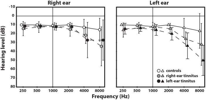
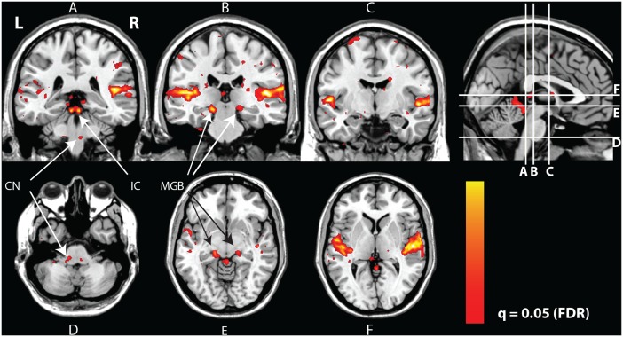
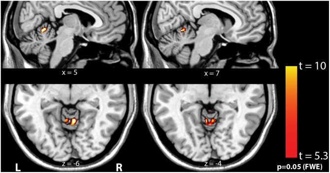
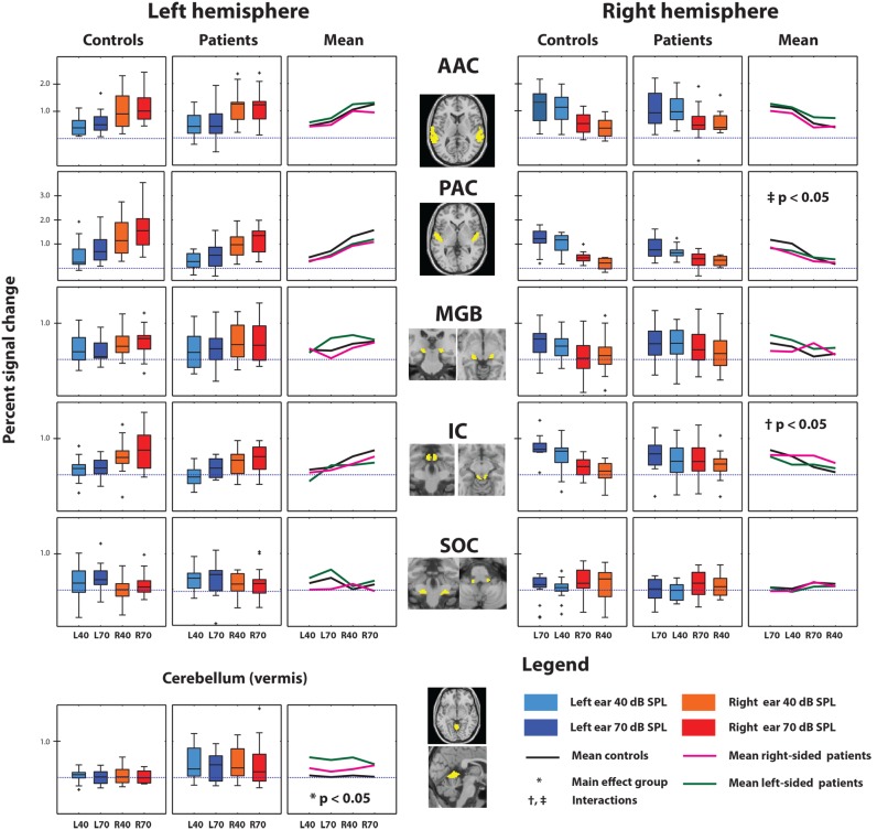
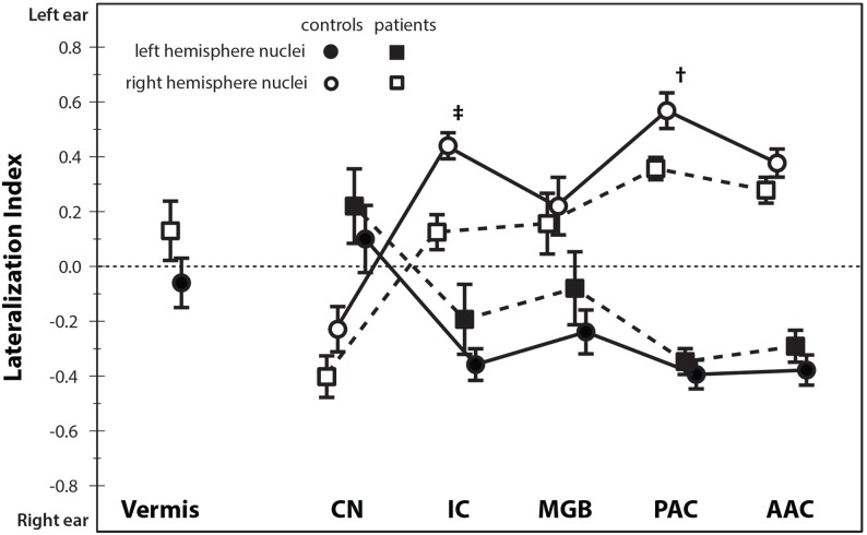
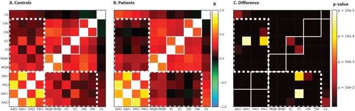
Tinnitus is a percept of sound that is not related to an acoustic source outside the body. For many forms of tinnitus, mechanisms in the central nervous system are believed to play a role in the pathology. In this work we specifically assessed possible neural correlates of unilateral tinnitus. Functional magnetic resonance imaging (fMRI) was used to investigate differences in sound-evoked neural activity between controls, subjects with left-sided tinnitus, and subjects with right-sided tinnitus. We assessed connectivity patterns between auditory nuclei and the lateralization of the sound-evoked responses. Interestingly, these response characteristics did not relate to the laterality of tinnitus. The lateralization for left- or right ear stimuli, as expressed in a lateralization index, was considerably smaller in subjects with tinnitus compared to that in controls, reaching significance in the right primary auditory cortex (PAC) and the right inferior colliculus (IC). Reduced functional connectivity between the brainstem and the cortex was observed in subjects with tinnitus. These differences are consistent with two existing models that relate tinnitus to i) changes in the corticothalamic feedback loops or ii) reduced inhibitory effectiveness between the limbic system and the thalamus. The vermis of the cerebellum also responded to monaural sound in subjects with unilateral tinnitus. In contrast, no cerebellar response was observed in control subjects. This suggests the involvement of the vermis of the cerebellum in unilateral tinnitus.
Conflict of interest statement
Figures
Similar articles
-
Reduced sound-evoked and resting-state BOLD fMRI connectivity in tinnitus.Neuroimage Clin. 2018 Aug 31;20:637-649. doi: 10.1016/j.nicl.2018.08.029. eCollection 2018. Neuroimage Clin. 2018. PMID: 30202725 Free PMC article.
-
Asymmetry in primary auditory cortex activity in tinnitus patients and controls.Neuroscience. 2014 Jan 3;256:117-25. doi: 10.1016/j.neuroscience.2013.10.015. Epub 2013 Oct 23. Neuroscience. 2014. PMID: 24161276
-
Tinnitus-related dissociation between cortical and subcortical neural activity in humans with mild to moderate sensorineural hearing loss.Hear Res. 2014 Jun;312:48-59. doi: 10.1016/j.heares.2014.03.001. Epub 2014 Mar 12. Hear Res. 2014. PMID: 24631963
-
Neural activity underlying tinnitus generation: results from PET and fMRI.Hear Res. 2009 Sep;255(1-2):1-13. doi: 10.1016/j.heares.2009.06.009. Epub 2009 Jun 21. Hear Res. 2009. PMID: 19545617 Review.
-
Auditory-limbic interactions in chronic tinnitus: Challenges for neuroimaging research.Hear Res. 2016 Apr;334:49-57. doi: 10.1016/j.heares.2015.08.005. Epub 2015 Aug 20. Hear Res. 2016. PMID: 26299843 Free PMC article. Review.
Cited by
-
Lateralization of cerebral blood flow in the auditory cortex of patients with idiopathic tinnitus and healthy controls: An arterial spin labeling study.Front Neurosci. 2022 Dec 7;16:992758. doi: 10.3389/fnins.2022.992758. eCollection 2022. Front Neurosci. 2022. PMID: 36636575 Free PMC article.
-
The association between subcortical and cortical fMRI and lifetime noise exposure in listeners with normal hearing thresholds.Neuroimage. 2020 Jan 1;204:116239. doi: 10.1016/j.neuroimage.2019.116239. Epub 2019 Oct 3. Neuroimage. 2020. PMID: 31586673 Free PMC article.
-
Aberrant thalamocortical coherence in an animal model of tinnitus.J Neurophysiol. 2019 Mar 1;121(3):893-907. doi: 10.1152/jn.00053.2018. Epub 2019 Jan 9. J Neurophysiol. 2019. PMID: 30625004 Free PMC article.
-
Altered functional connectivity of the thalamus in tinnitus patients is correlated with symptom alleviation after sound therapy.Brain Imaging Behav. 2020 Dec;14(6):2668-2678. doi: 10.1007/s11682-019-00218-0. Brain Imaging Behav. 2020. PMID: 31900891
-
Increased Resting-State Cerebellar-Cerebral Functional Connectivity Underlying Chronic Tinnitus.Front Aging Neurosci. 2018 Mar 5;10:59. doi: 10.3389/fnagi.2018.00059. eCollection 2018. Front Aging Neurosci. 2018. PMID: 29556191 Free PMC article.
References
-
- Noreña AJ, Eggermont JJ (2003) Changes in spontaneous neural activity immediately after an acoustic trauma: implications for neural correlates of tinnitus. Hear Res 183: 137–153. - PubMed
-
- Kaltenbach JA, Zacharek MA, Zhang J, Frederick S (2004) Activity in the dorsal cochlear nucleus of hamsters previously tested for tinnitus following intense tone exposure. Neurosci Lett 355: 121–125. - PubMed
-
- Seki S, Eggermont JJ (2003) Changes in spontaneous firing rate and neural synchrony in cat primary auditory cortex after localized tone-induced hearing loss. Hear Res 180: 28–38. - PubMed
Publication types
MeSH terms
LinkOut - more resources
Full Text Sources
Other Literature Sources
Medical
