

Intrauterine ischemic reperfusion switches the fetal transcriptional pattern from HIF-1α- to P53-dependent regulation in the murine brain
- PMID: 25329663
- PMCID: PMC4201554
- DOI: 10.1371/journal.pone.0110577
Intrauterine ischemic reperfusion switches the fetal transcriptional pattern from HIF-1α- to P53-dependent regulation in the murine brain
Abstract
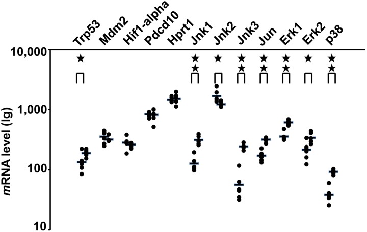
Ischemic reperfusion (IR) during the perinatal period is a known causative factor of fetal brain damage. So far, both morphologic and histologic evidence has shown that fetal brain damage can be observed only several hours to days after an IR insult has occurred. Therefore, to prevent fetal brain damage under these circumstances, a more detailed understanding of the underlying molecular mechanisms involved during an acute response to IR is necessary. In the present work, pregnant mice were exposed to IR on day 18 of gestation by clipping one side of the maternal uterine horn. Simultaneous fetal electrocardiography was performed during the procedure to verify that conditions resulting in fetal brain damage were met. Fetal brain sampling within 30 minutes after IR insult revealed molecular evidence that a fetal response was indeed triggered in the form of inhibition of the Akt-mTOR-S6 synthesis pathway. Interestingly, significant changes in mRNA levels for both HIF-1α and p53 were apparent and gene regulation patterns were observed to switch from a HIF-1α-dependent to a p53-dependent process. Moreover, pre-treatment with pifithrin-α, a p53 inhibitor, inhibited protein synthesis almost completely, revealing the possibility of preventing fetal brain damage by prophylactic pifithrin-α treatment.
Conflict of interest statement
Figures


Similar articles
-
Vaginal LPS changed gene transcriptional regulation response to ischemic reperfusion and increased vulnerability of fetal brain hemorrhage.Biochem Biophys Res Commun. 2015 Dec 4-11;468(1-2):228-33. doi: 10.1016/j.bbrc.2015.10.125. Epub 2015 Oct 30. Biochem Biophys Res Commun. 2015. PMID: 26523514
-
Isoflurane preconditioning protects rat brain from ischemia reperfusion injury via up-regulating the HIF-1α expression through Akt/mTOR/s6K activation.Cell Mol Biol (Noisy-le-grand). 2016 Feb 29;62(2):38-44. Cell Mol Biol (Noisy-le-grand). 2016. PMID: 26950449
-
Cilastatin Preconditioning Attenuates Renal Ischemia-Reperfusion Injury via Hypoxia Inducible Factor-1α Activation.Int J Mol Sci. 2020 May 19;21(10):3583. doi: 10.3390/ijms21103583. Int J Mol Sci. 2020. PMID: 32438631 Free PMC article.
-
Regulation of hypoxia-inducible factor-1α, regulated in development and DNA damage response-1 and mammalian target of rapamycin in human placental BeWo cells under hypoxia.Placenta. 2016 Sep;45:24-31. doi: 10.1016/j.placenta.2016.07.003. Epub 2016 Jul 14. Placenta. 2016. PMID: 27577706
-
Chemotherapy-mediated p53-dependent DNA damage response in clear cell renal cell carcinoma: role of the mTORC1/2 and hypoxia-inducible factor pathways.Cell Death Dis. 2013 Oct 17;4(10):e865. doi: 10.1038/cddis.2013.395. Cell Death Dis. 2013. PMID: 24136229 Free PMC article.
Cited by
-
Ultrasound Imaging of Mouse Fetal Intracranial Hemorrhage Due to Ischemia/Reperfusion.Front Physiol. 2017 May 24;8:340. doi: 10.3389/fphys.2017.00340. eCollection 2017. Front Physiol. 2017. PMID: 28596740 Free PMC article.
-
Amniotic LPS-Induced Apoptosis in the Fetal Brain Is Suppressed by Vaginal LPS Preconditioning but Is Promoted by Continuous Ischemic Reperfusion.Int J Mol Sci. 2022 Feb 4;23(3):1787. doi: 10.3390/ijms23031787. Int J Mol Sci. 2022. PMID: 35163709 Free PMC article.
-
Sulfated Polysaccharide Isolated from the Sea Cucumber Stichopus japonicus Against PC12 Hypoxia/Reoxygenation Injury by Inhibition of the MAPK Signaling Pathway.Cell Mol Neurobiol. 2015 Nov;35(8):1081-92. doi: 10.1007/s10571-015-0202-x. Epub 2015 May 8. Cell Mol Neurobiol. 2015. PMID: 25952102 Free PMC article.
-
Relationship Between Short Term Variability (STV) and Onset of Cerebral Hemorrhage at Ischemia-Reperfusion Load in Fetal Growth Restricted (FGR) Mice.Front Physiol. 2018 May 18;9:478. doi: 10.3389/fphys.2018.00478. eCollection 2018. Front Physiol. 2018. PMID: 29867536 Free PMC article.
References
-
- Carmeliet P, Dor Y, Herbert JM, Fukumura D, Brusselmans K, et al. (1998) Role of HIF-1α in hypoxia-mediated apoptosis, cell proliferation and tumour angiogenesis. Nature 394: 485–490. - PubMed
-
- Fandrey J (2004) Oxygen-dependent and tissue-specific regulation of erythropoietin gene expression. Am J Physiol Regul Integr Comp Physiol 286: R977–988. - PubMed
-
- Schmid P, Lorenz A, Hameister H, Montenarh M (1991) Expression of p53 during mouse embryogenesis. Development 113: 857–865. - PubMed
Publication types
MeSH terms
Substances
LinkOut - more resources
Full Text Sources
Other Literature Sources
Research Materials
Miscellaneous
