Case Reports
. 2014 Nov;64(11):569-75.
doi: 10.1111/pin.12207. Epub 2014 Oct 20.

Two autopsy cases of severe fever with thrombocytopenia syndrome (SFTS) in Japan: a pathognomonic histological feature and unique complication of SFTS

Affiliations
Free PMC article
Case Reports

Two autopsy cases of severe fever with thrombocytopenia syndrome (SFTS) in Japan: a pathognomonic histological feature and unique complication of SFTS

Tsubasa Hiraki et al. Pathol Int. 2014 Nov.
Free PMC article

Abstract

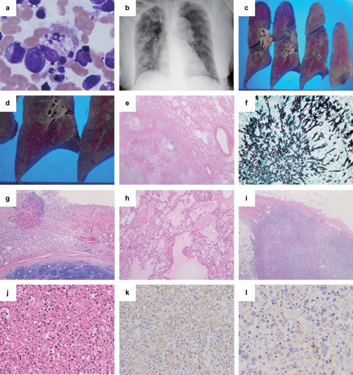
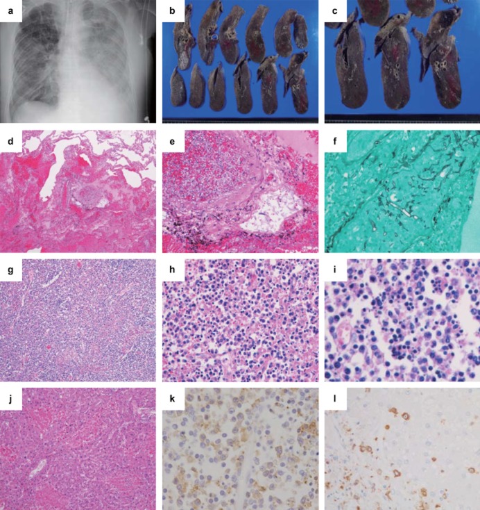
We report two autopsy cases of severe fever with thrombocytopenia syndrome (SFTS) with a high fatality rate in aged Japanese patients. Both cases were caused by a tick-bite. The pathognomonic histological feature was necrotizing lymphadenitis of systemic lymphoid tissue with SFTS viruses and SFTSV-RNA copies. Marked fungal infections were also observed in the lungs of both patients. Since cellular immune function may be suppressed in SFTS patients, physicians should be aware of possible fungal infections.

Keywords: SFTS; SFTS virus-nucleoprotein antigen; autopsy cases; fungal infection; necrotizing lymphadenitis.


Figures

Figure 1
Figure 1
Clinical images and pathological findings of Case 1. (a) Bone marrow finding and (b) chest X-ray image, (c,d) gross findings in the lungs, (e,g–j) hematoxylin and eosin staining, (f) Grocott staining, and (k,l) immunohistochemistry (IHC) using anti-SFTSV-NP antibody. (a) In the bone aspirate, many histiocytes show hemophagocytosis (×400). (b) A chest X-ray reveals a bilateral infiltrative shadow without consolidation. The cut surface of the right lung shows (c) many dispersed white nodular legions, (d) mainly in the lower lobe. (e) In the lung, there is necrotizing inflammation (×40) and (f)Aspergillus infection in the nodular lesions (Grocott staining ×400). (g) A tracheal ulcer with Aspergillus was also noted (×100). (h) Hyaline membrane formation indicating secondary diffuse alveolar damage is seen (×100). (i,j) In the left inguinal lymph node, the basic architecture of the lymph node is replaced by massive necrosis with infiltration of lymphocytes, histiocytes, some atypical lymphoid cells, and a significant amount of nuclear debris, but no neutrophils are observed (i, ×40; j, ×200). (k) In IHC of the lymph node, SFTSV-NP-positive cells are found (×100), and (l) positive staining for the SFTSV-NP antigen is detected in the cytoplasm of atypical lymphoid cells (×400).
Figure 2
Figure 2
Clinical images and pathological findings of case 2. (a) Chest X-ray image, (b,c) gross findings in the lungs, (d,e,g–j) hematoxylin and eosin staining, (f) Grocott staining, and (k,l) immunohistochemistry (IHC) using anti-SFTSV-NP antibody. (a) A chest X-ray reveals a bilateral infiltrative shadow without consolidation. (b,c) The cut surface of the right lung shows foci of pulmonary hemorrhage and infarction. (d) Diffuse hemorrhagic infarction (×40) and (e,f) angio-invasion of Mucor (e, ×200; f, Grocott staining ×400) are seen in the lung. (g,h) Necrotizing lymphadenitis is present in the lymph node around the abdominal aorta (g, ×40; h, ×400), and (i) hemophagocytosis is also observed (i, ×400). (j) The liver shows lobular necroses and mild portal fibrosis (×100). (k) IHC of the lymph node (×400) shows numerous SFTSV-NP-positive cells and positive signals for the SFTSV-NP antigen in the cytoplasm of atypical lymphoid cells. (l) IHC of the liver shows SFTSV-positive cells, but hepatocytes were negative for the SFTSV-NP antigen (×100).

References

    1. Yu XJ, Liang MF, Zhang SY, et al. Fever with thrombocytopenia associated with a novel bunyavirus in China. N Engl J Med. 2011;364:1523–1532. - PMC - PubMed
    1. Gai Z, Liang M, Zhang Y, et al. Person-to-person transmission of severe fever with thrombocytopenia syndrome bunyavirus through blood contact. Clin Infect Dis. 2012;54:249–252. - PMC - PubMed
    1. Liu Y, Li Q, Hu W, et al. Person-to-person transmission of severe fever with thrombocytopenia syndrome virus. Vector Borne Zoonotic Dis. 2012;12:156–160. - PubMed
    1. Tang X, Wu W, Wang H, et al. Human-to-human transmission of severe fever with thrombocytopenia syndrome bunyavirus through contact with infectious blood. J Infect Dis. 2013;207:736–739. - PubMed
    1. Takahashi T, Maeda K, Suzuki T, et al. The first identification and retrospective study of severe Fever with thrombocytopenia syndrome in Japan. J Infect Dis. 2013;209:816–827. - PMC - PubMed

Publication types

LinkOut - more resources