. 2014 Oct 17;9(10):e110296.
doi: 10.1371/journal.pone.0110296. eCollection 2014.

PLK1 and β-TrCP-dependent ubiquitination and degradation of Rap1GAP controls cell proliferation

Affiliations

PLK1 and β-TrCP-dependent ubiquitination and degradation of Rap1GAP controls cell proliferation

Dejie Wang et al. PLoS One. .

Abstract

Rap1GAP is a GTPase-activating protein (GAP) that specifically stimulates the GTP hydrolysis of Rap1 GTPase. Although Rap1GAP is recognized as a tumor suppressor gene and downregulated in various cancers, little is known regarding the regulation of Rap1GAP ubiquitination and degradation under physiological conditions. Here, we demonstrated that Rap1GAP is ubiquitinated and degraded through proteasome pathway in mitosis. Proteolysis of Rap1GAP requires the PLK1 kinase and β-TrCP ubiquitin ligase complex. We revealed that PLK1 interacts with Rap1GAP in vivo through recognition of an SSP motif within Rap1GAP. PLK1 phosphorylates Ser525 in conserved 524DSGHVS529 degron of Rap1GAP and promotes its interaction with β-TrCP. We also showed that Rap1GAP was a cell cycle regulator and that tight regulation of the Rap1GAP degradation in mitosis is required for cell proliferation.


Conflict of interest statement

Competing Interests: The authors have declared that no competing interests exist.

Figures

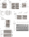
Figure 1
Figure 1. Rap1GAP is degraded during mitosis.
(A and B) HeLa cells were synchronized by double thymidine block followed by release into nocodazole-containing media and harvested at the indicated times. Cell cycle profile was assayed by FACS with propidium iodine staining (A). Protein levels were analyzed by immunoblotting (B). (C) Nocodazole-arrested HeLa cells were released into fresh medium, and the Rap1GAP status was monitored at indicated times. (D) HeLa cells were arrested using nocodazole at the indicated times, and 20 µM MG132 was added during the last 4 h of the nocodazole treatment before the cells were harvested. (E) HeLa cells or U2OS cells were treated with 20 µM MG132 and harvested at the indicated times. Protein levels were examined by immunoblotting. (F) HeLa cells that were synchronized by double thymidine block followed by release into nocodazole-containing media, and harvested at 22 h. The cell lysates were incubated with or without λ-phosphatase (λ-PPase). The phosphorylation state of Rap1GAP was analyzed by immunoblotting.
Figure 2
Figure 2. Rap1GAP is ubiquitinated and degraded in a β-TrCP dependent manner.
(A) 293T cells were co-transfected with Flag-Rap1GAP and the indicated Myc-tagged E3 ligases constructs (β-TrCP1, SKP2, Cdc20, Cdh1 and Fbw5). At 24 h after transfection, cells were harvested and lysed. Cell lysates were subjected to immunoprecipitation with anti-Myc antibody. The immunoprecipitates were immunoblotted with anti-Flag and anti-Myc antibodies, respectively. (B) 293T cells were co-transfected with HA-Rap1GAP and Flag- β-TrCP1/2 constructs. Cell lysates were prepared and subjected to immunoprecipitation with anti-HA or anti-Flag antibodies, respectively. (C) Endogenous Rap1GAP interacts with endogenous β-TrCP in HeLa cells. At the 4 h before harvesting, cells were treated with the proteasome inhibitor, MG132. HeLa cell lysates were incubated with protein A/G sepharose conjugated with either control IgG or Rap1GAP antibody. The immunoprecipitates were immunoblotted with Rap1GAP or β-TrCP antibodies, respectively. (D) Alignment of amino acids corresponding to the DSGxxS sequence with Rap1GAP1 orthologs and other β-TrCP substrates. (E) 293T cells were co-transfected with Flag-β-TrCP1/2 and wild type HA-Rap1GAP or SA mutant (S525A, S529A and S525/529A) constructs. At 24 h after transfection, cells were harvested and lysed. Cell lysates were subjected to immunoprecipitation with anti-HA antibody. The immunoprecipitates were immunoblotted with anti-Flag and anti-HA antibodies, respectively. (F) Flag-Rap1GAP (WT or S525/529A), HA-ubiquitin, and β-TrCP1 (WT or ΔF Box mutant) constructs were co-transfected into 293T cells. Rap1GAP proteins were immunoprecipitated by anti-Flag antibody. The polyubiquitinated forms of Rap1GAP were immunoblotted with anti-HA antibody. (G) Control siRNA or β-TrCP-specific siRNA were transfected in HeLa cells that were synchronized by double thymidine block followed by release into nocodazole-containing media for the indicated times. Cell lysates were subjected immunoblotting with the indicated antibodies. (H) Flag-Rap1GAP wild type or S525/529A mutant was transfected into HeLa cells that were synchronized by double thymidine block followed by release into nocodazole-containing media for the indicated times. Cell lysates were subjected to immunoblotting with the indicated antibodies.
Figure 3
Figure 3. PLK1 interacts with Rap1GAP and phosphorylates it at Ser525.
(A) 293T cells were co-transfected with Flag-Rap1GAP and Myc-PLK1 constructs. Cell lysates were prepared and subjected to immunoprecipitation with Flag or Myc antibodies, respectively. The immunoprecipitates were immunoblotted with anti-Flag and anti-Myc antibodies, respectively. (B) Endogenous Rap1GAP interacts with endogenous PLK1 in HeLa Cells. HeLa cells were synchronized by double thymidine block followed by release into nocodazole-containing media for 10 h. HeLa cells lysates were incubated with protein A/G sepharose conjugated with either control IgG or Rap1GAP antibody. The immunoprecipitates were immunoblotted with Rap1GAP or PLK1 antibodies, respectively. (C) Rap1GAP contains two PLK1 Polo box domain recognition motifs (S-S-P) located in the C-terminal sequence. (D) 293T cells were co-transfected with Myc-PLK1 and Flag-Rap1GAP (WT, S542A/P543A or S625A/P626A) (left panel), or HA-Rap1GAP (WT, S484A or S525/529A) constructs (right panel). Cell lysates were prepared and subjected to immunoprecipitation with Myc antibody. The immunoprecipitates were immunoblotted with anti-Flag and anti-HA antibodies, respectively. (E) Recombinant GST-Rap1GAP wild type or SA mutants (S525A, S529A and S525/529A) were phosphorylated by purified recombinant PLK1 wild type or kinase dead mutant (KD). Visualization by autoradiography (top panel) revealed Rap1GAP phosphorylation by PLK1. Coomasie brilliant blue staining (bottom panel) protein bands for Rap1GAP and PLK1.
Figure 4
Figure 4. PLK1 is essential for Rap1GAP degradation in mitosis.
(A) Control siRNA or PLK1-specific siRNA was transfected in Hela cells that were synchronized by double thymidine block followed by release into nocodazole-containing media for the indicated times. Cell lysates were subjected immunoblotting with the indicated antibodies. (B) HeLa cells were synchronized by double thymidine block followed by release into nocodazole-containing media for 15 h, then the cells were treated with the PLK1-specific inhibitor GW843682X (10 µM), GSK3β-specific inhibitor LiCl (10 mM), or control (DMSO) for indicated times, Cell lysates were subjected to immunoblotting with the indicated antibodies. (C) Flag-tagged Rap1GAP wild type or mutants (S484A, S625A/P626A) were transfected into HeLa cells that were synchronized by double thymidine block followed by release into nocodazole-containing media for the indicated times. Cell lysates were subjected to immunoblotting with the indicated antibodies.
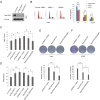
Figure 5
Figure 5. Rap1GAP degradation is required for cell proliferation.
(A) HeLa cells were transiently transfected with two Rap1GAP-specific siRNAs or control siRNA. At 48 h after transfection, cell lysates were subjected to immunoblotting with Rap1GAP antibody. (B) The cell cycle profile of Rap1GAP-knocked down HeLa cells were assayed by FACS with propidium iodine staining. (C and D) The proliferative capacity of Rap1GAP-knocked down or overexpressed HeLa (C) or U2OS (D) cells was measured by BrdU ELISA assay. HeLa cells or U2OS cells were transfected with Flag-Rap1GAP WT, S525/529A mutant or control constructs, the cell proliferative capacity were measured by BrdU ELISA assay. Similar ELISA assay was performed when Rap1GAP was knocked down. (E and F) HeLa (E) or U2OS (F) cells were transfected with Flag-Rap1GAP WT, S525/529A mutant or control constructs respectively and selected with G418 for 3 weeks and outgrowth colonies were stained by crystal violet. Representative photographs of cell colonies were shown. Total numbers of the colonies from three independent experiments were counted. *P<0.05; **, p<0.001.

Similar articles

Cited by

References

    1. Raaijmakers JH, Bos JL (2009) Specificity in Ras and Rap signaling. J Biol Chem 284: 10995–10999. - PMC - PubMed
    1. Bos JL, de Rooij J, Reedquist KA (2001) Rap1 signalling: adhering to new models. Nat Rev Mol Cell Biol 2: 369–377. - PubMed
    1. Zhang Z, Mitra RS, Henson BS, Datta NS, McCauley LK, et al. (2006) Rap1GAP inhibits tumor growth in oropharyngeal squamous cell carcinoma. Am J Pathol 168: 585–596. - PMC - PubMed
    1. Kim WJ, Gersey Z, Daaka Y (2012) Rap1GAP regulates renal cell carcinoma invasion. Cancer Lett 320: 65–71. - PMC - PubMed
    1. Zheng H, Gao L, Feng Y, Yuan L, Zhao H, et al. (2009) Down-regulation of Rap1GAP via promoter hypermethylation promotes melanoma cell proliferation, survival, and migration. Cancer Res 69: 449–457. - PubMed

Publication types

MeSH terms

Substances