. 2014 Oct 16;10(10):e1004448.
doi: 10.1371/journal.ppat.1004448. eCollection 2014 Oct.

Myosins VIII and XI play distinct roles in reproduction and transport of tobacco mosaic virus

Affiliations

Myosins VIII and XI play distinct roles in reproduction and transport of tobacco mosaic virus

Khalid Amari et al. PLoS Pathog. .

Abstract

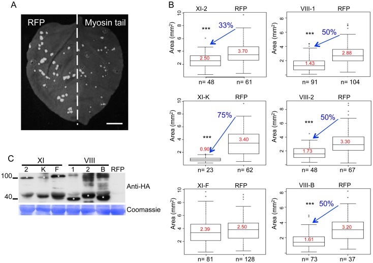
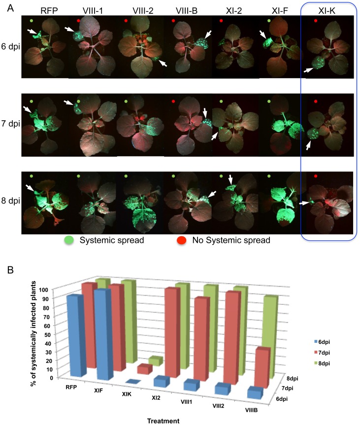
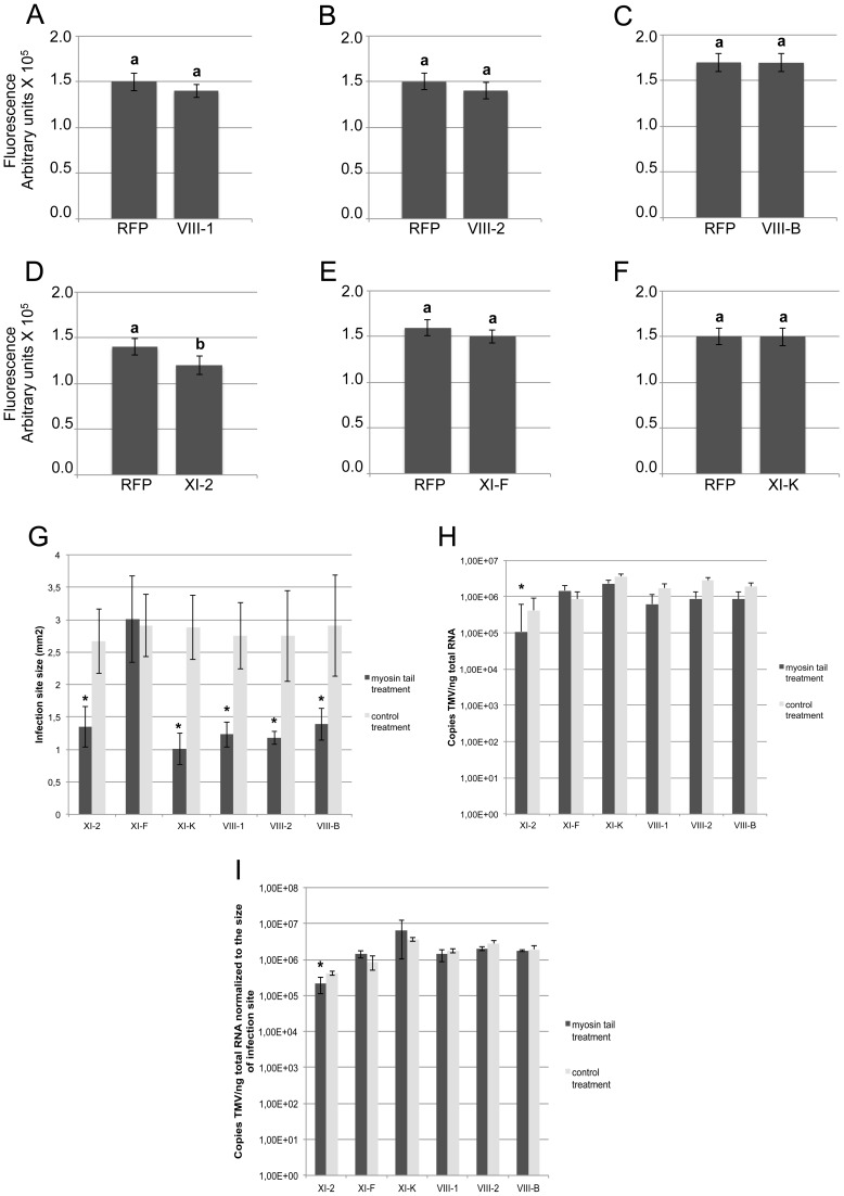
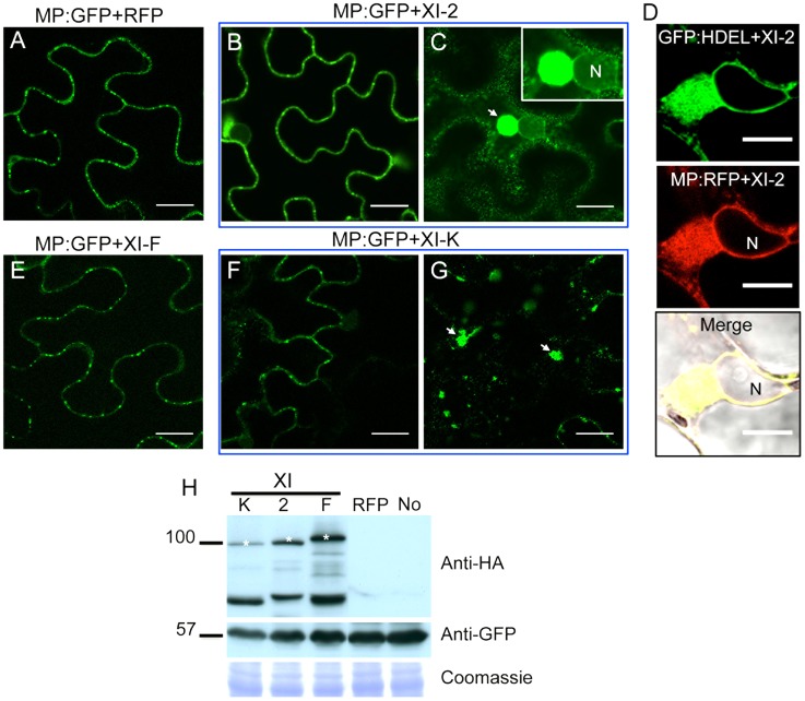
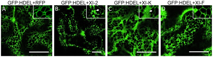
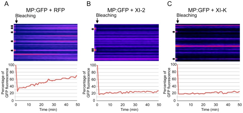
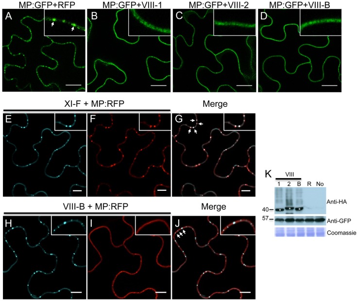
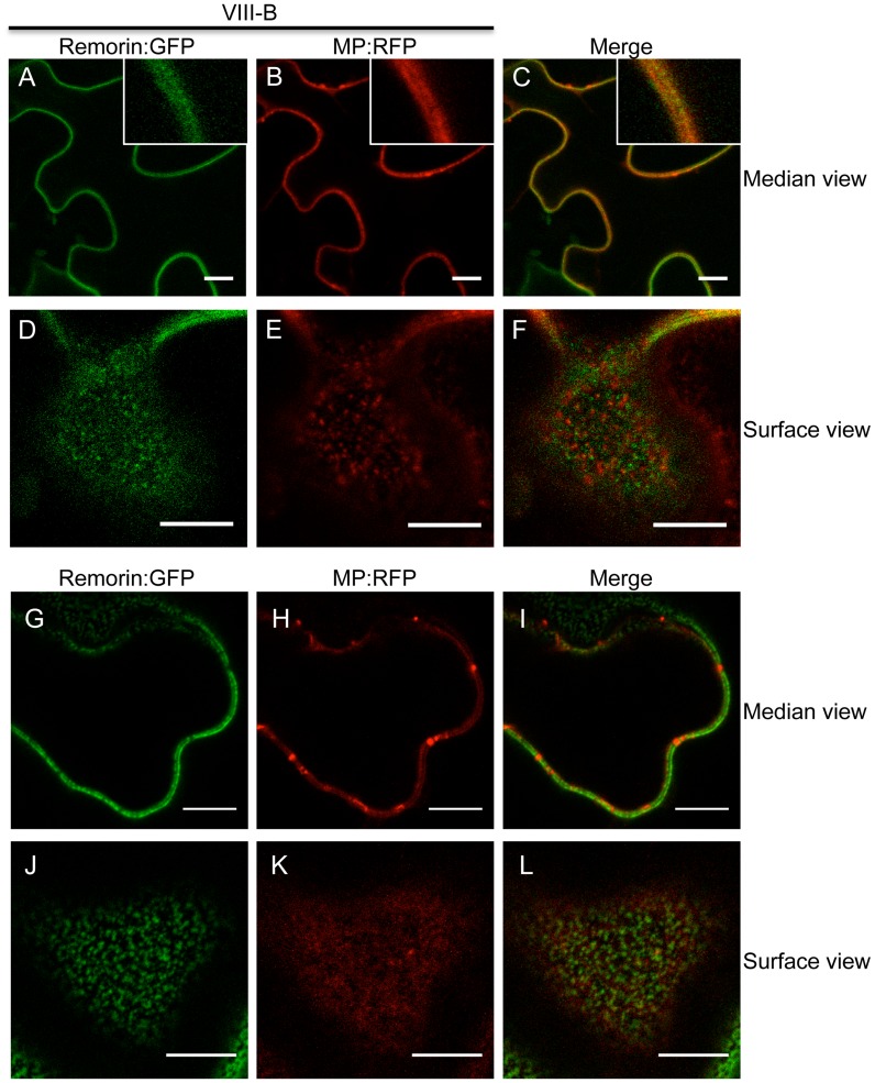
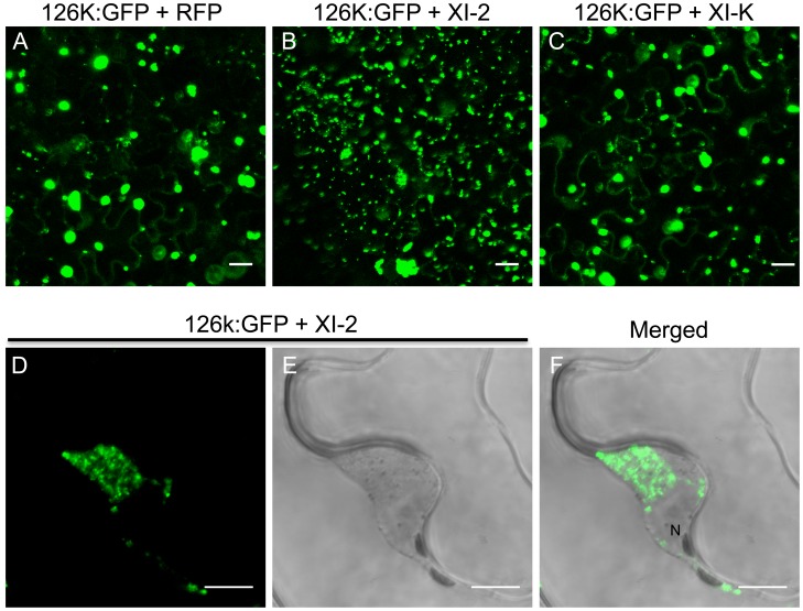
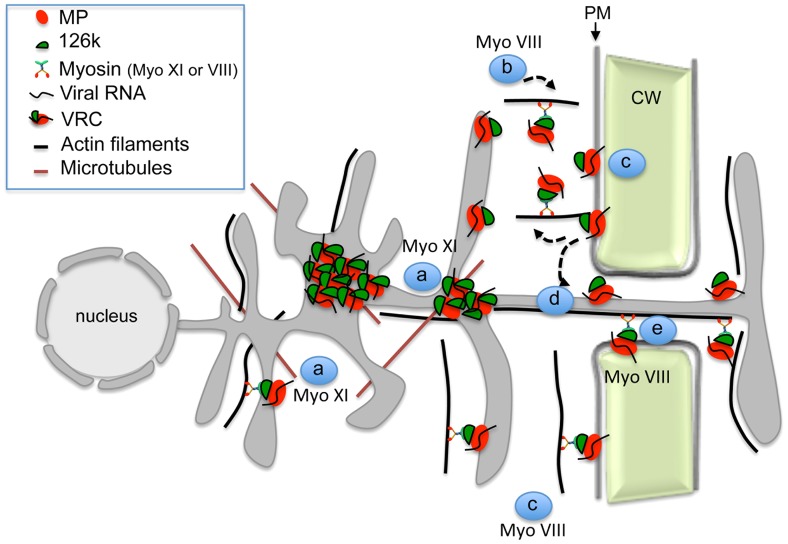
Viruses are obligatory parasites that depend on host cellular factors for their replication as well as for their local and systemic movement to establish infection. Although myosin motors are thought to contribute to plant virus infection, their exact roles in the specific infection steps have not been addressed. Here we investigated the replication, cell-to-cell and systemic spread of Tobacco mosaic virus (TMV) using dominant negative inhibition of myosin activity. We found that interference with the functions of three class VIII myosins and two class XI myosins significantly reduced the local and long-distance transport of the virus. We further determined that the inactivation of myosins XI-2 and XI-K affected the structure and dynamic behavior of the ER leading to aggregation of the viral movement protein (MP) and to a delay in the MP accumulation in plasmodesmata (PD). The inactivation of myosin XI-2 but not of myosin XI-K affected the localization pattern of the 126k replicase subunit and the level of TMV accumulation. The inhibition of myosins VIII-1, VIII-2 and VIII-B abolished MP localization to PD and caused its retention at the plasma membrane. These results suggest that class XI myosins contribute to the viral propagation and intracellular trafficking, whereas myosins VIII are specifically required for the MP targeting to and virus movement through the PD. Thus, TMV appears to recruit distinct myosins for different steps in the cell-to-cell spread of the infection.


Conflict of interest statement

The authors have declared that no competing interests exist.

Figures

Figure 1
Figure 1. Dominant negative repression of specific myosins inhibits TMV-GFP cell-to-cell spread.
A, example of an N. benthamiana leaf with TMV-GFP infection sites at 4 dpi in the presence of RFP in one half of the leaf and of inhibitory myosin VIII-1 tails in the other half. B, areas of viral infection sites are reduced in N. benthamiana half-leaves transiently expressing myosin VIII-1, VIII-2, VIII-B, XI-2 or XI-K tails as compared to half-leaves expressing RFP. Transient expression of myosin XI-F tails has no effect on the area of TMV-GFP viral infection sites. Boxplots depict the 25th to 75th percentile of measured areas of viral infection sites. Error bars indicate the range of the 5th and 95th percentile of measured areas of viral infection sites. The horizontal bar in each boxplot indicates the median value. Circles above the graphs represent outliers. n represents the number of viral infection sites measured on three half-leaves. Mean area of viral infection sites is given in red. (***) represents significant differences as determined by unbalanced ANOVA (p<0.001). Data are from three independent experiments. C, immunoblot analysis using HA- (top panel) antibodies revealed the expression of the HA-tagged myosin tails. Bands corresponding to class XI (≈100 kDa) and class VIII (≈40 kDa) myosin tails are marked by asterisks. Coomassie blue staining (bottom panel) is shown as loading control. All leaves were inoculated with equal volumes of TMV-GFP in-vitro transcription reaction mix and agro-infiltrated for myosin tail/RFP expression two days later. Analyses were conducted at 4 dpi.
Figure 2
Figure 2. Transient expression of specific myosin tails in the inoculated leaf delays or inhibits the onset of systemic spread of TMV-GFP-JL24.
A, the onset of systemic spread is delayed upon transient expression of myosin tails XI-2, VIII-B, VIII-1 and VIII-2 in the inoculated leaf (arrow), whereas expression of XI-F tails has no effect. Overexpression of myosin tails XI-K inhibits the systemic movement of TMV. Images are representative for the results obtained in three independent experiments, each with four plants for each treatment. Single leaves of each plant were inoculated with equal amounts of TMV-GFP-JL24 virions and agroinfiltrated for myosin tail/RFP expression two days later. Plants were scored for systemic infection at 6, 7 and 8 dpi. B, percentage of systemically infected plants at 6, 7 and 8 dpi.
Figure 3
Figure 3. Transient expression of myosin tail XI-2, but not the expression of any of the other tested myosin tails, affects virus replication.
A-F, Fluorescence intensity (arbitrary units) of the TMV-GFP infection sites analyzed in Figure 1. Fluorescence intensity was quantified using ImageJ software and normalized to the area of the same infection sites. Except for myosin XI-2 tails (Figure 3D), myosin tail expression had no significant effect on the fluorescence intensity of infection sites (Figure 3A-C, E and F). Different letters (a and b) above the columns indicate statistically significant differences between the different treatments, as determined by t-test (P<0.001). G-I, quantification of TMV-GFP RNA upon overexpression of myosin tails. Plants were inoculated with equal amount of TMV-GFP and agro-infiltrated for expression with myosin tails/RFP two days later. Analyses of infection sites were conducted at 4 dpi. At least 38 infection sites from three different leaves taken from three different plants was analysed per treatment. G, size of TMV-GFP infection sites upon expression of myosin tails compared to the control (RFP). With exception of myosin XI-F tails, the expression of all other tested myosin tails significantly reduced the area of the infection sites. H, quantification of TMV-GFP RNA accumulation within individual infection sites upon myosin tail expression. For all treatments, the same amount of leaf material carrying three infection sites was used for total RNA extraction. The statistical analysis is based on three independent experiments (three biological replicates). Overexpression of myosin XI-2 tails reduces the accumulation of TMV-GFP, whereas the expression of all other test myosin tails had no effect. I, copies of TMV-GFP genomes as shown in Figure 3H normalized to the area of the respective infection sites in Figure 3G. Among the tested myosin tails, only myosin XI-2 tails affect viral RNA accumulation. Statistical significance was determined by t-test (* = P<0.05). Bars represent means and standard errors.
Figure 4
Figure 4. Transient expression of myosin XI-2 and XI-K tails changes the accumulation pattern of the MP.
A, co-expression of MP:GFP with RFP. The MP:GFP accumulates in PD. B and C, co-expression of the MP:GFP together with myosin XI-2 tails does not affect significantly the localization of the MP:GFP to PD (B) but causes the accumulation of the MP:GFP in a single big aggregate in the vicinity of the nucleus (C, arrow). D, transient co-expression of myosin XI-2 tails and MP:RFP in N. benthamiana transgenic plants expressing GFP:HDEL as ER marker (16c). Accumulation of the MP:RFP in a single aggregate near to the nucleus upon inactivation of myosin XI-2 correlates with the accumulation of the GFP:HDEL indicating that the MP aggregates are ER-derived. E, co-expression of the MP:GFP with myosin XI-F tails. The MP:GFP accumulates in PD similar as in the control with RFP (A). F and G, co-expression of the MP:GFP together with myosin XI-K tails has no significant effect on the localization of the MP:GFP to PD (F) but causes accumulation of the MP:GFP in several aggregates (G, arrow). H, immunoblot analysis using HA-specific (top panel) and GFP-specific (middle panel) antibodies for the detection of myosin and MP:GFP, respectively. Bands corresponding to the class XI (≈100 kDa) myosin tails are marked by asterisks. Coomassie blue staining (bottom panel) is shown as loading control. A-H, proteins were expressed by co-agroinfiltration and observed at 1 dpa. N, Nucleus. No, MP:GFP expressed alone. Scale bars, 20 µm (A-C and E-G) and 10 µm (D).
Figure 5
Figure 5. Effect of specific myosin tail expression on ER structure.
A, transient expression of RFP in 16c plants at 2 dpa. ER network structure is not affected and the normal tubular ER structure is seen. B, expression of myosin XI-2 tails in 16c plants. The tubular structure of the ER is disrupted and GFP-containing aggregates are formed (arrow inside the inset). C, expression of myosin XI-K tails in 16c plants. The normal tubular structure of the ER is inhibited. Instead, large sheet-like membrane structures are seen (arrow inside the inset). D, expression of myosin XI-F tails in 16c plants. No effect on the ER structure is observed. A-D, proteins were expressed by co-agroinfiltration and observed at 1 dpa. Scale bars, 20 µm.
Figure 6
Figure 6. Inhibition of myosin XI-2 and XI-K reduces the efficiency by which MP is targeted to PD.
A-C, Kyomographs showing examples of fluorescence recovery during the time of the FRAP experiments (50 min, from the left to the right) after bleaching of several PD (arrows) at 1 dpa. A, FRAP experiment in the presence of RFP. MP:GFP fluorescence at photobleached PD recovers to 70% during 50 minutes after bleaching (lower panel). B, FRAP experiment in the presence of myosin XI-2 tails. MP:GFP fluorescence at PD recovers only to 30% during 50 minutes (lower panel). C, FRAP experiment in the presence of myosin XI-K tails. Like in the presence of XI-2, MP:GFP fluorescence at PD recovers only to 30% during 50 minutes (lower panel). Fat arrows at the left side of the kymographs indicate the location of photobleached PD. Arrows on the top of the kymographs indicate the bleaching time point.
Figure 7
Figure 7. Inactivation of myosin VIII-1, VIII-2 or VIII-B disrupts the PD localization of MP.
A, expression of RFP. The expression of RFP does not affect the accumulation of the co-expressed MP:GFP in PD (arrow inside the inset). B-D, expression of myosin VIII tails. The presence of either myosin VIII-1 tails (B), myosin VIII-2 tails (C), or of myosin VIII-B tails (D) interferes with the localization of MP:GFP to PD. E to G, myosin XI-F tail expression does not interfere with the co-localization of MP:RFP with aniline blue-stained callose at PD. E, aniline blue staining of callose; F, MP:RFP; G, merged images. H to J, myosin VIII-B tail expression interferes with the co-localization of MP:RFP with aniline blue-stained callose at PD. H, aniline blue staining of callose; I, MP:RFP; J, merged images. K, immunoblot analysis using HA-specific (top panel) and GFP-specific (middle panel) antibodies for the detection of myosin and MP:GFP, respectively. Bands corresponding to the class VIII (≈40 kDa) myosin tails are marked by asterisks. Coomassie blue staining (bottom panel) is shown as loading control. R, MP:GFP expressed with RFP. No, MP:GFP expressed alone. A-K, proteins were expressed by co-agroinfiltration and analyzed at 1 dpa. Scale bars, 20 µm (A-D); 10 µm (E-J).
Figure 8
Figure 8. Inactivation of myosin VIII causes patchy accumulation of MP at the PM.
A to C, remorin:GFP and MP:RFP co-localize at the PM in the presence of myosin VIII-B tails. A, remorin:GFP; B, MP:RFP; C, merged images. D to F, surface view on the PM. Remorin:GFP and MP:RFP localize to different patches at the PM in the presence of myosin VIII-B tails. D, remorin:GFP; E, MP:RFP; F, merged images. G to I, in the absence of myosin VIII-B tails the MP:RFP accumulates in PD. G, remorin:GFP; H, MP:RFP; I, merged images. J to L, surface view of the PM. In the absence of myosin VIII-B tails, the MP:RFP does not accumulate in patchy pattern within the PM. J, remorin:GFP; K, MP:RFP; L, merged images. Proteins were expressed by co-agroinfiltration and observed at 1 dpa. Scale bars, 10 µm.
Figure 9
Figure 9. Inhibition of myosin XI-2 disrupts the normal subcellular localization of expressed 126k:GFP.
A, co-expression of 126k:GFP together with RFP. The 126k:GFP accumulates in large aggregates. B, co-expression of 126k:GFP together with myosin XI-2 tails. 126k:GFP is dispersed into numerous small aggregates. C, co-expression of 126k:GFP together with myosin XI-K tails. The 126k:GFP accumulates in large aggregates similar to those seen upon expression of RFP. Similar normal aggregates are seen also upon co-expression of the 126k:GFP with either XI-F, VIII-1, VIII-2 or VIII-B tails. D-F, co-expression of the 126k:GFP together with myosin XI-2 tails. The 126k:GFP accumulates, in addition to small aggregates, in a big aggregate in the vicinity of the nucleus. A-F, proteins were expressed by co-agroinfiltration and observed at 2 dpa. Scale bars, 20 µm (A-C); 10 µm (D-F).
Figure 10
Figure 10. Model illustrating the role of the actomyosin system in the intra- and intercellular movement of TMV.
Myosins XI-2 and XI-K provide motility to the ER and facilitate the concentration of VRCs at cortical ER sites in the vicinity of the PM (a). VRCs are concentrated and stabilized by the 126k through myosin XI-2 function to enhance viral replication and silencing suppression (a). Class VIII myosins are involved in the targeting of the MP/VRCs from the cortical ER sites to the PM and subsequently to PD. This process may involve endocytic recycling (b), diffusion of the MP/VRC along the PM (c), stabilization of MP/VRC at PD (d) and active transport through the channel into the adjacent cell (e).

References

    1. Burch-Smith TM, Zambryski PC (2012) Plasmodesmata paradigm shift: regulation from without versus within. Annu Rev Plant Biol 63: 239–260. - PubMed
    1. Maule A, Faulkner C, Benitez-Alfonso Y (2012) Plasmodesmata "in Communicado". Front Plant Sci 3: 30. - PMC - PubMed
    1. Niehl A, Heinlein M (2011) Cellular pathways for viral transport through plasmodesmata. Protoplasma 248: 75–99. - PubMed
    1. Amari K, Boutant E, Hofmann C, Schmitt-Keichinger C, Fernandez-Calvino L, et al. (2010) A family of plasmodesmal proteins with receptor-like properties for plant viral movement proteins. PloS Pathog 6: e1001119. - PMC - PubMed
    1. Amari K, Lerich A, Schmitt-Keichinger C, Dolja VV, Ritzenthaler C (2011) Tubule-guided cell-to-cell movement of a plant virus requires class XI myosin motors. PLoS Pathog 7: e1002327. - PMC - PubMed

Publication types

MeSH terms

LinkOut - more resources