Review
. 2015 May 1;22(13):1111-29.
doi: 10.1089/ars.2014.5994. Epub 2015 Jan 19.

Redox regulation of NLRP3 inflammasomes: ROS as trigger or effector?

Affiliations
Review

Redox regulation of NLRP3 inflammasomes: ROS as trigger or effector?

Justine M Abais et al. Antioxid Redox Signal. .

Abstract

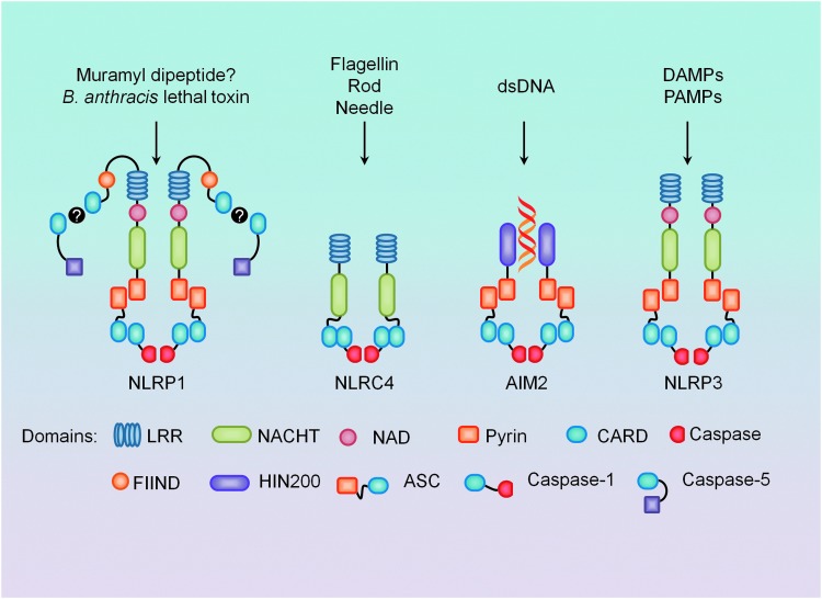
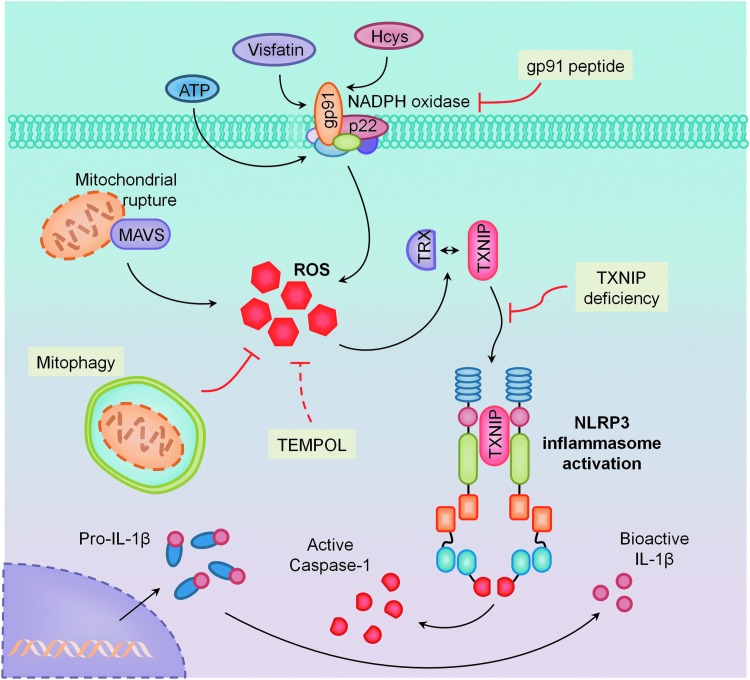
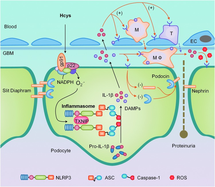
Significance: Inflammasomes are multiprotein complexes localized within the cytoplasm of the cell that are responsible for the maturation of proinflammatory cytokines such as interleukin-1β (IL-1β) and IL-18, and the activation of a highly inflammatory form of cell death, pyroptosis. In response to infection or cellular stress, inflammasomes are assembled, activated, and involved in host defense and pathophysiology of diseases. Clarification of the molecular mechanisms leading to the activation of this intracellular inflammatory machinery may provide new insights into the concept of inflammation as the root of and route to human diseases.

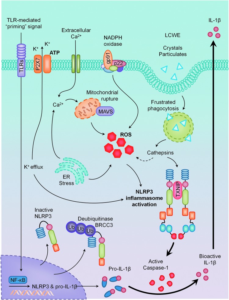
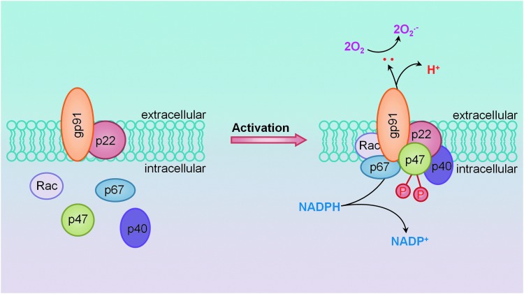
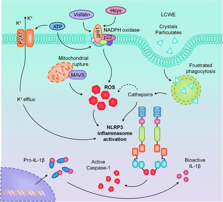
Recent advances: The activation of inflammasomes, specifically the most fully characterized inflammasome-the nucleotide-binding oligomerization domain (NOD)-like receptor containing pyrin domain 3 (NLRP3) inflammasome, is now emerging as a critical molecular mechanism for many degenerative diseases. Several models have been developed to describe how NLRP3 inflammasomes are activated, including K(+) efflux, lysosome function, endoplasmic reticulum (ER) stress, intracellular calcium, ubiquitination, microRNAs, and, in particular, reactive oxygen species (ROS).

Critical issues: ROS may serve as a "kindling" or triggering factor to activate NLRP3 inflammasomes as well as "bonfire" or "effector" molecules, resulting in pathological processes. Increasing evidence seeks to understand how this spatiotemporal action of ROS occurs during NLRP3 inflammasome activation, which will be a major focus of this review.

Future directions: It is imperative to know how this dual action of ROS works during NLRP3 inflammation activation on different stimuli and what relevance such spatiotemporal redox regulation of NLRP3 inflammasomes has in cell or organ functions and possible human diseases.


Figures

<b>FIG. 1.</b>
FIG. 1.
Four major types of inflammasomes and their known stimulators. Nucleotide-binding oligomerization domain (NOD)-like receptor containing pyrin domain 3 (NLRP1), proposed to be activated by bacterial cell wall component muramyl dipeptide (MDP) and Bacillus anthracis lethal toxin, can directly cause caspase-5 processing, but the presence of adaptor protein apoptosis-associated speck-like protein containing a CARD (ASC) results in more robust activation. NLRC4 activation is mostly associated with gram-negative bacteria components and can also directly process caspase-1 through its caspase recruitment domain (CARD). Double-stranded DNA (dsDNA) binds preferentially to the HIN200 domain of AIM2 (absent in melanoma 2), and requires ASC for caspase-1 processing. NLRP3 also requires ASC and caspase-1, is activated in response to both exogenous and endogenous danger signals, and is mostly recognized for its role in sterile inflammation. To see this illustration in color, the reader is referred to the web version of this article at www.liebertpub.com/ars
<b>FIG. 2.</b>
FIG. 2.
Models of NLRP3 inflammasome activation. Considered to be a two-step mechanism, the primary signal comes from the activation of toll-like receptors (TLRs) and is responsible for the upregulation of NLRP3 and pro-interleukin-1β (IL-1β) in an NF-kappaB (NF-κB)-dependent manner. Secondary signals come from multiple pathways: K+ efflux via P2X7 receptor activation, endoplasmic reticulum (ER) stress, mitochondrial dysfunction, NADPH oxidase, frustrated phagocytosis, and lysosomal rupture pathways, all of which appear to converge in the production of reactive oxygen species (ROS). Together, these primary and secondary signals activate the NLRP3 inflammasome, resulting in proteolytic cleavage of caspase-1 and the maturation of IL-1β. To see this illustration in color, the reader is referred to the web version of this article at www.liebertpub.com/ars
<b>FIG. 3.</b>
FIG. 3.
Membrane NADPH oxidase assembly and activation through lipid raft (LR)-mediated clustering to form redox signaling platforms. Under rest condition, all subunits of NADPH oxidase are separated and the enzyme may not be active. When vascular or kidney cells are stimulated by inflammatory stimuli such as Hcys or visfatin, the formation of LR platforms occurs. In such platforms, NADPH oxidase subunits such as gp91phox and p47phox and other proteins become aggregated, clustered, or recruited, resulting in a rapid assembling of NADPH oxidase into an enzyme complex, producing O2•− and ROS that conduct transmembrane or intracellular signaling. To see this illustration in color, the reader is referred to the web version of this article at www.liebertpub.com/ars
<b>FIG. 4.</b>
FIG. 4.
Primary activating pathways of NLRP3 inflammasomes. It was demonstrated that in the kidney and vasculature, NLRP3 inflammasomes are activated by NADPH oxidase-derived ROS through LR clustering. Some stimuli such as ATP also alter K+ efflux or lysosome stability to activate NLRP3 inflammasomes. To see this illustration in color, the reader is referred to the web version of this article at www.liebertpub.com/ars
<b>FIG. 5.</b>
FIG. 5.
Mediators of ROS action to activate NLRP3 inflammasomes. Two distinct proteins have been demonstrated to be associated with NLRP3–thioredoxin-interacting protein (TXNIP) and mitochondrial anti-viral signaling protein (MAVS). TXNIP as a binding partner to NLRP3 time dependently dissociates from thioredoxin (TRX) and then binds with NLRP3, leading to inflammasome formation and activation. MAVS also associates with NLRP3 to facilitate its oligomerization with ASC and caspase-1. To see this illustration in color, the reader is referred to the web version of this article at www.liebertpub.com/ars
<b>FIG. 6.</b>
FIG. 6.
Implications of NLRP3 inflammasomes in podocyte injury and ultimate glomerular sclerosis. In response to pathological stimuli, NADPH oxidase in podocytes or glomerular endothelial cells is activated via LR clustering to produce O2•−, which results in the formation of NLRP3 inflammasomes to produce IL-1β, IL-18, and other molecules such as damage-associated molecular patterns (DAMPs). These factors may recruit and activate inflammatory cells such as macrophages (MΦ) and T-cells in glomeruli, where “bonfire” O2•− and cytokines are produced to initiate typical chronic sterile glomerular inflammation, leading to tissue injury and sclerosis. Coinciding with other direct actions of IL-1β or other inflammasome products, this sterile inflammatory response leads to podocyte loss and foot process effacement, progressing into glomerular sclerosis and end-stage renal disease (ESRD). To see this illustration in color, the reader is referred to the web version of this article at www.liebertpub.com/ars
<b>FIG. 7.</b>
FIG. 7.
The formation and activation of NLRP3 inflammasome in the endothelium of carotid arteries during ligation injury. The formation and activation of endothelial NLRP3 inflammasomes were observed in the intima of carotid arteries during ligation that induces vascular injury or inflammation locally. IL-1β production in the intima could almost be completely blocked by caspase-1 inhibitor and ASC gene silencing or knocking out. Scale bar=10 μm. To see this illustration in color, the reader is referred to the web version of this article at www.liebertpub.com/ars

References

    1. Abais JM, Xia M, Boini KM, and Li PL. Contribution of guanine nucleotide exchange factor Vav2 to homocysteine-induced NLRP3 inflammasome activation in mouse podocytes. FASEB J 28: 1063.6, 2014
    1. Abais JM, Xia M, Li G, Chen Y, Conley SM, Gehr TW, Boini KM, and Li PL. Nod-like receptor protein 3 (NLRP3) inflammasome activation and podocyte injury via thioredoxin-interacting protein during hyperhomocysteinemia. J Biol Chem 289: 27159, 2014 - PMC - PubMed
    1. Abais JM, Xia M, Li G, Gehr TW, Boini KM, and Li PL. Contribution of endogenously produced reactive oxygen species to the activation of podocyte NLRP3 inflammasomes in hyperhomocysteinemia. Free Radic Biol Med 67: 211–220, 2014 - PMC - PubMed
    1. Abais JM, Zhang C, Xia M, Liu Q, Gehr TW, Boini KM, and Li PL. NADPH oxidase-mediated triggering of inflammasome activation in mouse podocytes and glomeruli during hyperhomocysteinemia. Antioxid Redox Signal 18: 1537–1548, 2013 - PMC - PubMed
    1. Aganna E, Martinon F, Hawkins PN, Ross JB, Swan DC, Booth DR, Lachmann HJ, Bybee A, Gaudet R, Woo P, Feighery C, Cotter FE, Thome M, Hitman GA, Tschopp J, and McDermott MF. Association of mutations in the NALP3/CIAS1/PYPAF1 gene with a broad phenotype including recurrent fever, cold sensitivity, sensorineural deafness, and AA amyloidosis. Arthritis Rheum 46: 2445–2452, 2002 - PubMed

Publication types

MeSH terms