. 2015 Jan 15;465(2):325-35.
doi: 10.1042/BJ20141069.

Structural determinants of host specificity of complement Factor H recruitment by Streptococcus pneumoniae

Affiliations

Structural determinants of host specificity of complement Factor H recruitment by Streptococcus pneumoniae

David Achila et al. Biochem J. .

Abstract

Many human pathogens have strict host specificity, which affects not only their epidemiology but also the development of animal models and vaccines. Complement Factor H (FH) is recruited to pneumococcal cell surface in a human-specific manner via the N-terminal domain of the pneumococcal protein virulence factor choline-binding protein A (CbpAN). FH recruitment enables Streptococcus pneumoniae to evade surveillance by human complement system and contributes to pneumococcal host specificity. The molecular determinants of host specificity of complement evasion are unknown. In the present study, we show that a single human FH (hFH) domain is sufficient for tight binding of CbpAN, present the crystal structure of the complex and identify the critical structural determinants for host-specific FH recruitment. The results offer new approaches to the development of better animal models for pneumococcal infection and redesign of the virulence factor for pneumococcal vaccine development and reveal how FH recruitment can serve as a mechanism for both pneumococcal complement evasion and adherence.


Figures
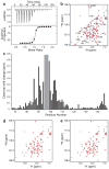
Figure 1
Figure 1. ITC and NMR analysis of the binding of hFH and CbpA
a, ITC measurement of the binding of hFH CCP9. For the ITC experiment, the syringe was loaded with a 75 μM solution of CbpAN and the sample cell with 5 μM hFH CCP9. The injections were made over a period of 120 min with a 6 min interval between subsequent injections. The sample cell was stirred at 310 rpm. b, Overlaid NMR spectra of CbpAN (blue) and CbpAN + CCP9 (red). The 1H-15N HSQC NMR spectra were obtained with 1.0 mM 15N-CbpAN in the absence and the presence of 1.1 mM hFH CCP9. Complete sequential resonance assignments are presented in Supplementary Figure 1. c, Perturbation of the chemical shifts of the backbone amides of CbpAN by the binding of hFH CCP9. The loop region with missing assignment for the free protein is indicated by a gray bar. Chemical shift changes were calculated using the equation: Δδ = {0.5[Δδ(1HN)2 + (0.2Δδ(15N))2 }1/2. d, Overlaid NMR spectra of CbpAN (blue) and CbpAN + CCP13-15 (red). The 1H-15N HSQC NMR spectra were obtained with 0.2 mM 15N-CbpAN in the absence and the presence of 0.5 mM CCP13-15. e, Overlaid NMR spectra of CbpAN (blue) and CbpAN + CCP19-20 (red). The 1H-15N HSQC NMR spectra were obtained with 0.2 mM 15N-CbpAN in the absence and the presence of 1.0 mM CCP19-20.
Figure 2
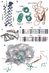
Figure 2. Structural analysis of CbpAN and its complex with hFH CCP9
a, NMR structure of free CbpAN. The Cα traces of the top 20 conformers are drawn. CbpAN has a compact structure consisting of a bundle of three helices with an up-down-up antiparallel topology. b, Crystal structure of the complex of CbpAN (green) and hFH CCP9 (orange) superposed with the NMR structure of free CbpAN (cyan). The side-chains that form the two disulfide bonds (S-S) are drawn in purple lines. c, Superposed crystal structures of hFH CCP9 (blue), CCP4 (red), and CCP8 (green). Cα RMSDs between CCP9 and CCP4 (PDB ID: 2WII) and between CCP9 and CCP8 (PDB ID: 2V8E) are 1.139 Å for 54 residues and 1.344 Å for 59 residues, respectively. d, Structure-based amino acid sequence alignment of hFH CCP9, CCP4, and CCP8. The conserved residues between the three CCPs are shaded in black and CCP9 residues at the interface of binding CbpAN are colored in red. The numbering is that of CCP9. e, Hydrogen bonding and hydrophobic locking between hFH CCP9 and CbpAN. hFH residues are lettered in orange, CbpA residues are lettered in cyan, and hydrogen bonds are indicated by dash lines. The hydrophobic lock (a cluster of hydrophobic residues, Val 495, Met 497, Leu 543 and Ile 545) of hFH CCP9 is drawn in a surface representation.
Figure 3
Figure 3. Amino acid sequence alignment of CCP9s from 18 mammals
The residues conserved among the 18 CCP9s are shaded in black. The interface residues of hFH CCP9 in the binding of CbpAN are indicated with ◆ and the residues that constitute the “hydrophobic lock” with ■. The residues of mFH CCP9 substituted with the corresponding residues of hFH CCP9 in the mutagenesis study are indicated with #. The abbreviations for various mammals are: NOMLE, Nomascus leucogenys (Northern white-cheeked gibbon); PONAB, Pongo abelii (Sumatran orangutan); MACFA, Macaca fascicularis (crab-eating macaque); MACMU, Macaca mulatta (Rhesus macaque); CALJA, Callithrix jacchus (white-tufted-ear marmoset); LOXAF, Loxodonta africana (African elephant); HETGA, Heterocephalus glaber (naked mole rat); OTOGA, Otolemur garnettii (small-eared galago); AILME, Ailuropoda melanoleuca (giant panda).
Figure 4
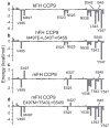
Figure 4. Computational analysis of the binding of CCP9 and CbpAN
Binding energy decompositions were obtained by MM-GBSA analysis for the wild-type hFH CCP9 (a), the hFH CCP9 triple mutant protein with Met 497 replaced by Glu, Leu 543 by Thr, and Ile 545 by Ser (b), the wild-type mFH CCP9 (c), and the mFH CCP9 triple mutant protein with Glu 497 replaced by Met, Thr 543 by Leu, and Ser 545 by Ile (d).

References

    1. Ricklin D, Hajishengallis G, Yang K, Lambris JD. Complement: a key system for immune surveillance and homeostasis. Nat Immunol. 2010;11:785–797. - PMC - PubMed
    1. Serruto D, Rappuoli R, Scarselli M, Gros P, van Strijp JAG. Molecular mechanisms of complement evasion: learning from staphylococci and meningococci. Nature Rev Microbiol. 2010;8:393–399. - PubMed
    1. Ferreira VP, Pangburn MK, Cortes C. Complement control protein factor H: The good, the bad, and the inadequate. Mol Immunol. 2010;47:2187–2197. - PMC - PubMed
    1. Zipfel PF, Skerka C. Complement regulators and inhibitory proteins. Nat Rev Immunol. 2009;9:729–740. - PubMed
    1. Granoff DM, Welsch JA, Ram S. Binding of Complement Factor H (fH) to Neisseria meningitidis Is Specific for Human fH and Inhibits Complement Activation by Rat and Rabbit Sera. Infect Immun. 2009;77:764–769. - PMC - PubMed

Publication types

MeSH terms