. 2015 Jan;33(1):91-100.
doi: 10.1002/stem.1869.

Regulated apoptosis of genetically modified hematopoietic stem and progenitor cells via an inducible caspase-9 suicide gene in rhesus macaques

Affiliations

Regulated apoptosis of genetically modified hematopoietic stem and progenitor cells via an inducible caspase-9 suicide gene in rhesus macaques

Cecilia N Barese et al. Stem Cells. 2015 Jan.

Abstract

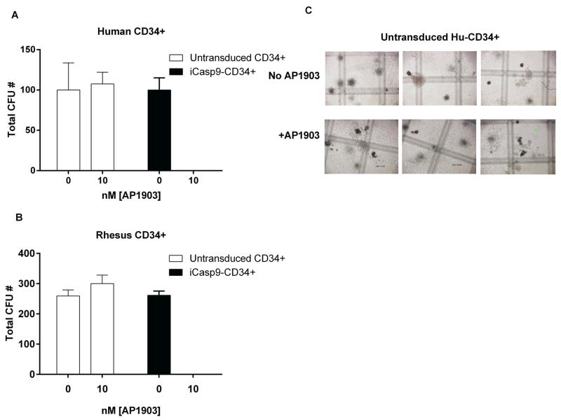
The high risk of insertional oncogenesis reported in clinical trials using integrating retroviral vectors to genetically modify hematopoietic stem and progenitor cells (HSPCs) requires the development of safety strategies to minimize risks associated with novel cell and gene therapies. The ability to ablate genetically modified cells in vivo is desirable, should an abnormal clone emerge. Inclusion of "suicide genes" in vectors to facilitate targeted ablation of vector-containing abnormal clones in vivo is one potential safety approach. We tested whether the inclusion of the "inducible Caspase-9" (iCasp9) suicide gene in a gamma-retroviral vector facilitated efficient elimination of vector-containing HSPCs and their hematopoietic progeny in vivo long-term, in an autologous non-human primate transplantation model. Following stable engraftment of iCasp9 expressing hematopoietic cells in rhesus macaques, administration of AP1903, a chemical inducer of dimerization able to activate iCasp9, specifically eliminated vector-containing cells in all hematopoietic lineages long-term, suggesting activity at the HSPC level. Between 75% and 94% of vector-containing cells were eliminated by well-tolerated AP1903 dosing, but lack of complete ablation was linked to lower iCasp9 expression in residual cells. Further investigation of resistance mechanisms demonstrated upregulation of Bcl-2 in hematopoietic cell lines transduced with the vector and resistant to AP1903 ablation. These results demonstrate both the potential and the limitations of safety approaches using iCasp9 to HSPC-targeted gene therapy settings, in a model with great relevance to clinical development.

Keywords: Gene therapy; Genotoxicity; HSC transplantation; Suicide gene; iCasp9.


Conflict of interest statement

No other authors have relevant conflicts of interest.

Figures

Fig. 1
Fig. 1. Effect of AP1903 on human or rhesus CD34+ cells transduced with the iCasp suicide vector
A. Summary of % cell death of human CD34+ cells as determined by staining for Annexin V and 7ADD (% death includes both 7ADD + and Ann-V+ cells). Human CD34+ cells transduced with the SFG-iCasp9-2A-ΔCD19 vector (black) and sorted for ΔCD19 expression, are compared with CD34+ cells that were not transduced (white), or CD34+ cells transduced with a control MND-MFG-GFP vector (no iCasp9 gene) (grey). Cells were exposed overnight to AP1903 before analysis. Values shown are mean ± SEM. Two-way ANOVA was used for analyses of significance. The p values of 10 nM and 50 nM AP1903 % cell death for SFG-iCasp9-2A-ΔCD19-transduced cells compared to either the untransduced or control vector-transduced cells were all **** < 0.0001; n= 4. There was no significant impact of AP1903 on cell death of untransduced or control vector-transduced CD34+ cells, comparing 0 to 10 or 50 nM AP1903. B. Representative dot plot panel showing flow cytometric analysis of human CD34+ cells transduced with the SFG-iCasp9-2A-ΔCD19 vector and then exposed to AP1903 overnight, and stained for Annexin-V and 7AAD apoptotic markers. C. % cell death (mean ± SEM) for rhesus CD34+ cells transduced with SFG-iCasp9-2A-ΔCD19 and exposed to AP1903, compared to untransduced rhesus CD34+ cells. % cell death was significantly increased between untransduced and transduced cells exposed to 10 nM (***p= 0.001) and 50 nM (***p= 0.001), respectively, and between transduced untreated and transduced treated with 10 nM and 50 nM AP1903 (****p= 0.0001; n=2). There was no significant difference in cell death between untransduced cells incubated with 0 versus 10 nM or 50 nM AP1903, nor between untransduced untreated and transduced untreated rhesus CD34+ cells (2-way ANOVA). D. Representative panel showing flow cytometric analysis of animal A7X028 CD34+ cells transduced with the SFG-iCasp9-2A-ΔCD19 vector and then exposed to AP1903 overnight, and stained for Annexin-V and 7AAD apoptotic markers.
Fig. 2
Fig. 2. Colony forming units (CFU) assays on human and rhesus CD34+ cells transduced with the SFG-iCasp9-2A-.CD19 vector
A. Human, and B. rhesus CD34+ cells were transduced with the vector, sorted for ΔCD19, and treated or not treated with 10nM AP1903 overnight before plating in CFU assays. C. In parallel, untransduced Hu and Rh CD34+ cells were incubated overnight with 10 and 50 nM AP1903 and plated. CFU were enumerated at day 15. Abbreviations: ns (p= > 0.05; n=3)
Fig. 3
Fig. 3. SFG-iCasp9-2A-ΔCD19 transduction and autologous bone marrow transplantation in rhesus macaques
A. Summary of transduction efficiency and cell transplantation doses for three rhesus macaques transplanted with autologous CD34+ transduced with the SFG-iCasp9-2A-ΔCD19 vector. B. Marking levels in vivo as assessed by flow cytometry analysis for ΔCD19 expression in peripheral blood leukocytes of the three macaques at different time points after transplantation. C. The effect of the AP1903 therapy on the level of ΔCD19+ cells in myeloid (CD18+ and CD14+), T (CD3+), and B (CD20+) peripheral blood lineages, as assessed by flow cytometry. Levels of ΔCD19+ cells before (white bars) and 6 months after (dark bars) the last dose of AP1903 are shown. D. Flow cytometric histograms of ΔCD19 expression in PB and BM of animal A7X028 at 9 months after transplant prior to AP1903 treatment, and 45 days after AP1903 discontinuation. E. Longitudinal analysis of the level of ΔCD19+ cells as assessed by flow cytometry in macaques transplanted with SFG-iCasp9-2A-ΔCD19-transduced autologous CD34+ cells. The timelines indicates days post initial treatment with AP1903. The “PreAP1903” time point was 10 months in DCLZ and A7X028 and 6 months in A7E065, and levels were completely stable in all animals at these time points (see panel 3B above). Infusions of 0.4 mg/Kg (light gray arrows), 1.4 mg/Kg (dark gray arrows) and 4 mg/Kg (black arrows) AP1903 are indicated. F. Vector copy number as assessed by real time-quantitative PCR in PB leukocytes before initiation of AP1903 therapy and ~ 45 days after AP1903 discontinuation (right panel). The flow cytometric analyses of CD19 expresion level in PB is also presented (left panel).
Fig. 4
Fig. 4. Analyses of the characteristics of residual CD19+ cells after AP1903 therapy
A. Mean fluorescence intensity (MFI) for ΔCD19 in peripheral blood leukocytes from macaques transplanted with autologous transduced CD34+ cells before and after completion of AP1903. B. PBMCs from AP1903-treated macaques were cultured overnight with and without IL-2 and anti-CD3/CD28 beads and with and without AP1903. After overnight incubation the cells were assayed by flow cytomety for the expression of ΔCD19. Means of duplicate assays for each sample are shown. C. Karpas 299 cells transduced with the iCasp9-ΔCD19 vector were sorted for high (black) and low (grey) hCD19 expression and then exposed to 50nM AP1903 for 3 consecutive days in vitro (grey and black arrows indicate results after each day of in vitro dosing). Data represent means ± SEM; n=3. Abbreviations: ns= p >0.05; *** p= 0.0008. D. Western blots for expression of the cleaved fragment of caspase 9 (37 kDa) and caspase 3 (17/19 kDa) in Karpas cells transduced with SFG-iCasp9-ΔCD19 with and without exposure to AP1903 overnight. Results from the entire cell population as well as sorted ΔCD19+ cells are shown. E. Western blots for expression of uncleaved caspase 9 (47 kDa) in untransduced and transduced Karpas 299 cells. The transduced cells were sorted for high and low ΔCD19 expression by flow cytometry. Band intensities on triplicate determination normalized for actin expression are reported as mean ± SEM in the bar graphs below the blots. F. Western blot determination of expression of XIAP (53 kDa) and Bcl2 (28 kDa) in control untransduced Karpas cells, and transduced hCD19+ Karpas cells resistant to AP1903. The control and resistant cells (in duplicate) were re-exposed to 50 nM AP1903, overnight.

References

    1. Hacein-Bey-Abina S, Garrigue A, Wang GP, Soulier J, Lim A, Morillon E, Clappier E, Caccavelli L, Delabesse E, Beldjord K, Asnafi V, MacIntyre E, Dal Cortivo L, Radford I, Brousse N, Sigaux F, Moshous D, Hauer J, Borkhardt A, Belohradsky BH, Wintergerst U, Velez MC, Leiva L, Sorensen R, Wulffraat N, Blanche S, Bushman FD, Fischer A, Cavazzana-Calvo M. Insertional oncogenesis in 4 patients after retrovirus-mediated gene therapy of scid-x1. J Clin Invest. 2008;118:3132–3142. - PMC - PubMed
    1. Hacein-Bey-Abina S, Von Kalle C, Schmidt M, McCormack MP, Wulffraat N, Leboulch P, Lim A, Osborne CS, Pawliuk R, Morillon E, Sorensen R, Forster A, Fraser P, Cohen JI, de Saint Basile G, Alexander I, Wintergerst U, Frebourg T, Aurias A, Stoppa-Lyonnet D, Romana S, Radford-Weiss I, Gross F, Valensi F, Delabesse E, Macintyre E, Sigaux F, Soulier J, Leiva LE, Wissler M, Prinz C, Rabbitts TH, Le Deist F, Fischer A, Cavazzana-Calvo M. Lmo2-associated clonal t cell proliferation in two patients after gene therapy for scid-x1. Science. 2003;302:415–419. - PubMed
    1. Howe SJ, Mansour MR, Schwarzwaelder K, Bartholomae C, Hubank M, Kempski H, Brugman MH, Pike-Overzet K, Chatters SJ, de Ridder D, Gilmour KC, Adams S, Thornhill SI, Parsley KL, Staal FJ, Gale RE, Linch DC, Bayford J, Brown L, Quaye M, Kinnon C, Ancliff P, Webb DK, Schmidt M, von Kalle C, Gaspar HB, Thrasher AJ. Insertional mutagenesis combined with acquired somatic mutations causes leukemogenesis following gene therapy of scid-x1 patients. The Journal of clinical investigation. 2008;118:3143–3150. - PMC - PubMed
    1. Stein S, Ott MG, Schultze-Strasser S, Jauch A, Burwinkel B, Kinner A, Schmidt M, Kramer A, Schwable J, Glimm H, Koehl U, Preiss C, Ball C, Martin H, Gohring G, Schwarzwaelder K, Hofmann WK, Karakaya K, Tchatchou S, Yang R, Reinecke P, Kuhlcke K, Schlegelberger B, Thrasher AJ, Hoelzer D, Seger R, von Kalle C, Grez M. Genomic instability and myelodysplasia with monosomy 7 consequent to evi1 activation after gene therapy for chronic granulomatous disease. Nature medicine. 2010;16:198–204. - PubMed
    1. Riviere I, Dunbar CE, Sadelain M. Hematopoietic stem cell engineering at a crossroads. Blood. 2012;119:1107–1116. - PMC - PubMed

Publication types