. 2014 Nov 21;34(6):e00152.
doi: 10.1042/BSR20140120.

The putative signal peptide of glucagon-like peptide-1 receptor is not required for receptor synthesis but promotes receptor expression

Affiliations

The putative signal peptide of glucagon-like peptide-1 receptor is not required for receptor synthesis but promotes receptor expression

Yunjun Ge et al. Biosci Rep. .

Abstract

GLP-1R (glucagon-like peptide-1 receptor) mediates the 'incretin effect' and many other anti-diabetic actions of its cognate ligand, GLP-1 (glucagon-like peptide-1). It belongs to the class B family of GPCRs (G protein-coupled receptors) and possesses an N-terminal putative SP (signal peptide). It has been reported that this sequence is required for the synthesis of GLP-1R and is cleaved after receptor synthesis. In the present study, we conducted an in-depth exploration towards the role of the putative SP in GLP-1R synthesis. A mutant GLP-1R without this sequence was expressed in HEK293 cells (human embryonic kidney 293 cells) and displayed normal functionality with respect to ligand binding and activation of adenylate cyclase. Thus the putative SP does not seem to be required for receptor synthesis. Immunoblotting analysis shows that the amount of GLP-1R synthesized in HEK293 cells is low when the putative SP is absent. This indicates that the role of the sequence is to promote the expression of GLP-1R. Furthermore, epitopes tagged at the N-terminal of GLP-1R are detectable by immunofluorescence and immunoblotting in our experiments. In conclusion, the present study points to different roles of SP in GLP-1R expression which broadens our understanding of the functionality of this putative SP of GLP-1R and possibly other Class B GPCRs.


Figures
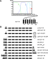
Figure 1
Figure 1. Signal peptide prediction and cDNA construct illustration
(A) Depiction of the putative signal sequence and the cleavage site. N-terminal amino acids of GLP-1R were monitored with the program ‘SignalP 3.0’. The probabilities of the presence of n (green), h (blue) and c (light blue) regions, and the cleavage probabilities (cp, red) are indicated in a score ranging from 0 to 1. (B) Presentation of the constructs used in the present study. The SPs and the transmembrane domains are shown as gray or black boxes. N-tail sequences are indicated as open boxes. HA and FLAG tags are shown as grey boxes. Fused GFP is indicated. Signal peptides of GLP-1R, CRF1R and CRF2(a)R are the first 23, 24 and 19 amino acids, respectively.
Figure 2
Figure 2. Pharmacological properties of the mutant GLP-1R lacking the putative SP with the wild-type receptor as a control
(A) Binding curves for increasing concentrations of the unlabelled ligand GLP-1 (21.4 pM–166.7 nM) to displace binding of the radiolabelled ligand 125I-GLP-1 (200 pM) to HEK293 cells transfected with wild-type and mutant receptors. Values are calculated as percentages of maximal specific binding obtained in the presence of 21.4 pM unlabelled GLP-1. Non-specific binding and maximal specific binding of the mutant are similar to those of the wild-type (two-tailed paired t test). (B) Curves for intracellular cAMP responses in HEK293 cells to stimulation by increasing concentrations of GLP-1 (0.1 pM–10 nM). Values are calculated as percentages of maximal cAMP responses stimulated by 10 nM GLP-1. Basal and maximal cAMP levels of the mutant are similar to those of the wild-type (two-tailed paired t test). Data shown are means±S.E.M. from three independent experiments with each performed in triplicates.
Figure 3
Figure 3. Comparison of GLP-1R-GFP and ΔSP-GLP-1R-GFP expression
(A) Confocal laser scanning microscopy of HEK293 cells transiently expressing GLP-1R-GFP and ΔSP-GLP-1R-GFP with wild-type GLP-1R as a control. GFP intensity is shown in green (left panel) and membrane stained with CellMask™ Deep Red is displayed in red (central panel). Merge of both is exhibited in yellow (right panel). Horizontal scanning of representative cells is shown. Scale bar=10 μm. (B) Immunoblotting of GLP-1R expression and glycosylation in transiently transfected HEK293 cells. Lysates of cells expressing GLP-1R-GFP and ΔSP-GLP-1R-GFP were treated with Endo H or PNGase F to remove glycans or left untreated. Receptor proteins were detected with rabbit anti-GFP and HRP-linked goat anti-rabbit antibodies. Each lane shows a protein load from 5×10⁵ cells with β-actin as a control (detected with mouse anti-β-actin antibodies). The image is a representative of three independent experiments. (C) mRNA measurements were made in HEK293 cells transiently transfected with GLP-1R-GFP or ΔSP-GLP-1R-GFP. Relative mRNA levels were depicted as fold difference in mRNA expression after being normalized to the reference sample (cells expressing the wild-type GLP-1R) with GAPDH as an internal control. Data shown are means±S.E.M. from three independent experiments with each performed in triplicates. No statistically significant difference was found (two-tailed paired t test).
Figure 4
Figure 4. Detection of HA epitopes tagged at N/C-termini of different receptors expressed in HEK293 cells
(A) Confocal laser scanning microscopy after immunostaining intact cells. HA-CRF1R, HA-CRF2(a)R and HA-GLP-1R constructs were transiently transfected and receptors at the cell surfaces were labelled with mouse anti-HA antibodies and Alexa Fluor® 488 donkey anti-mouse antibodies (left and right panels, green). Signal of Hochest 33342 staining is shown in blue (right panel). Horizontal scanning of representative cells is shown. Scale bar=10 μm. (B) Immunoblotting of lysates from HEK293 cells expressing wild-type GLP-1R, HA-GLP-1R and GLP-1R-HA. Receptor proteins were detected with mouse anti-HA and HRP-linked secondary anti-mouse antibodies. Each lane shows receptor proteins from 5×10⁵ cells with β-actin as a control. The immunoblotting shown is representative of three independent experiments.
Figure 5
Figure 5. Immunoblotting analysis of the impact of epitope tagging on GLP-1R expression
The indicated constructs were transiently expressed in HEK293 cells and the wild-type GLP-1R was used as a control. Receptor proteins were detected with mouse anti-HA antibodies (left panel) or mouse anti-FLAG antibodies (right panel) plus HRP-linked secondary anti-mouse antibodies. Samples for HA-GLP-1R-FLAG and GLP-1R-FLAG-HA are shown as HA-GLP-1RF and GLP-1RF-HA, respectively. Each lane shows receptor proteins from 5×10⁵ cells with β-actin as a control. Immunoblotting displayed is representative of three independent experiments.
Figure 6
Figure 6. Immunofluorescence microscopy and immunoblotting analyses with the double-labelled GLP-1R
(A) The constructs, HSF-GLP-1R and SF-GLP-1R, were transiently expressed in HEK293 cells and immunofluorescence microscopy was performed as described above. Signal of Alexa Fluor® 488 donkey anti-mouse antibodies is shown in green and that of Hochest 33342 staining in blue. Horizontal scanning of representative cells is shown. Scale bar=10 μm. (B) The indicated constructs were transiently expressed in HEK293 cells and wild-type GLP-1R was used as a control. Receptor proteins were detected with mouse anti-HA antibodies (left panel) or mouse anti-FLAG antibodies (right panel) by immunoblotting. Each lane shows receptor proteins from 5×10⁵ cells with β-actin as a control. Immunoblotting displayed is representative of three independent experiments.

Similar articles

Cited by

References

    1. Willard F. S., Sloop K. W. Physiology and emerging biochemistry of the glucagon-like peptide-1 receptor. Exp. Diabetes Res. 2012;2012:470851. - PMC - PubMed
    1. Leech C. A., Dzhura I., Chepurny O. G., Kang G., Schwede F., Genieser H. G., Holz G. G. Molecular physiology of glucagon-like peptide-1 insulin secretagogue action in pancreatic beta cells. Prog. Biophys. Mol. Biol. 2011;107:236–247. doi: 10.1016/j.pbiomolbio.2011.07.005. - DOI - PMC - PubMed
    1. Willard F. S., Bueno A. B., Sloop K. W. Small molecule drug discovery at the glucagon-like peptide-1 receptor. Exp. Diabetes Res. 2012;2012:1–9. - PMC - PubMed
    1. Kochl R., Alken M., Rutz C., Krause G., Oksche A., Rosenthal W., Schulein R. The signal peptide of the G protein-coupled human endothelin B receptor is necessary for translocation of the N-terminal tail across the endoplasmic reticulum membrane. J. Biol. Chem. 2002;277:16131–16138. doi: 10.1074/jbc.M111674200. - DOI - PubMed
    1. Schulein R., Westendorf C., Krause G., Rosenthal W. Functional significance of cleavable signal peptides of G protein-coupled receptors. Eur. J. Cell Biol. 2012;91:294–299. doi: 10.1016/j.ejcb.2011.02.006. - DOI - PubMed

Publication types

LinkOut - more resources