. 2014 Dec 15;53(51):14096-14101.
doi: 10.1002/anie.201408533. Epub 2014 Oct 21.

PET imaging of bacterial infections with fluorine-18-labeled maltohexaose

Affiliations

PET imaging of bacterial infections with fluorine-18-labeled maltohexaose

Xinghai Ning et al. Angew Chem Int Ed Engl. .

Abstract

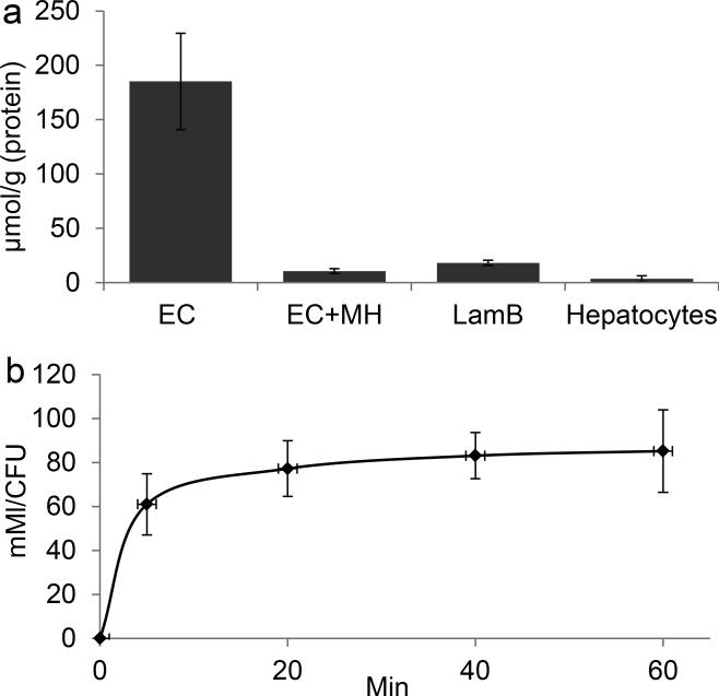
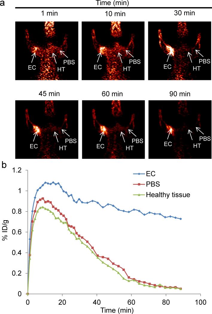
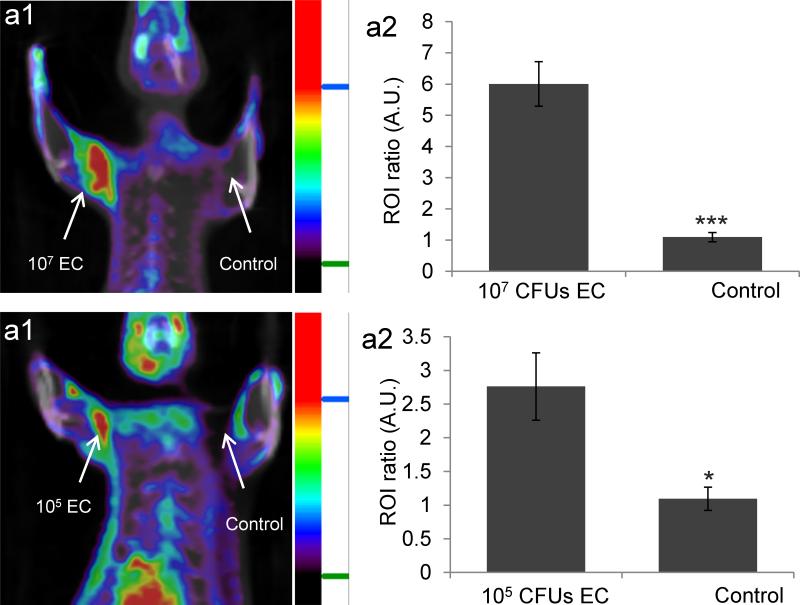
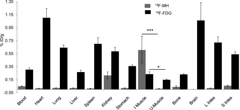
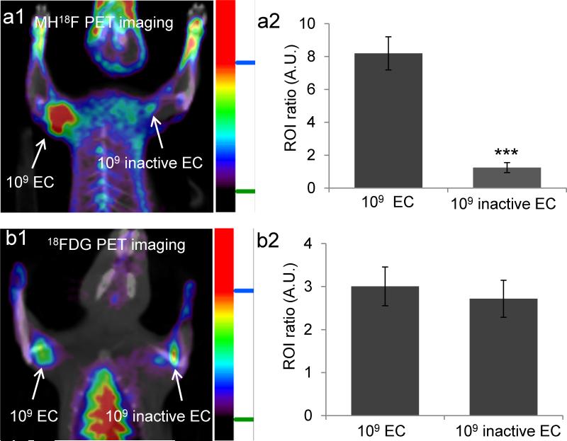
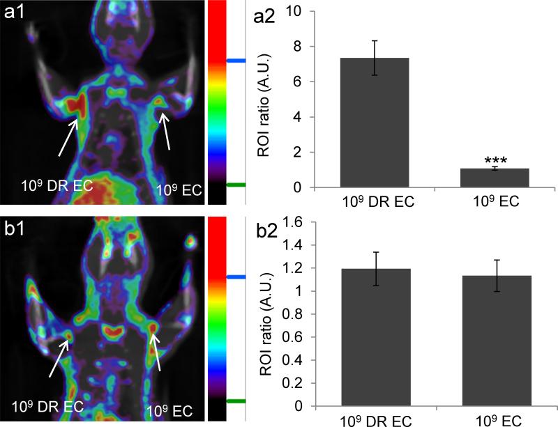
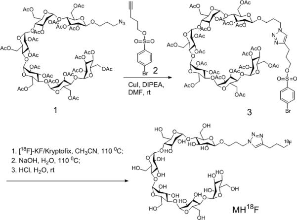
A positron emission tomography (PET) tracer composed of (18)F-labeled maltohexaose (MH(18)F) can image bacteria in vivo with a sensitivity and specificity that are orders of magnitude higher than those of fluorodeoxyglucose ((18)FDG). MH(18)F can detect early-stage infections composed of as few as 10(5) E. coli colony-forming units (CFUs), and can identify drug resistance in bacteria in vivo. MH(18)F has the potential to improve the diagnosis of bacterial infections given its unique combination of high specificity and sensitivity for bacteria.

Keywords: bacterial infections; maltodextrin transporters; maltohexaose; positron emission tomography; radiochemistry.


Figures

Figure 1
Figure 1
MH19F has high specificity for bacteria and is robustly internalized by bacteria. a, MH19F has high specificity for bacteria over hepatocytes. E. coli (EC), EC with LamB mutation (LamB) and mammalian cells were incubated with 500 μM MH19F for 1 hour in the presence or absence of 50 mM maltohexaose (MH). The intracellular MH19F concentration was determined and normalized to protein content. Bacteria robustly accumulate MH19F whereas hepatocytes have negligible uptake. The uptake of MH19F in EC is inhibited by a large excess of maltohexaose, and the uptake of MH19F in LamB mutants is significantly reduced. The results are expressed as mean micromoles per gram of protein ± s.e.m. for n = 3 per group. b, The accumulation of MH19F in EC reaches millimolar concentrations. EC were incubated with 500 μM MH19F, and the intracellular concentration of MH19F was determined at different time points, n = 3 per group.
Figure 2
Figure 2
In vivo PET imaging of rats infected with E.coli (107CFUs). a. Rats were infected in the left triceps muscle with 107 E.coli, injected with MH18F, and dynamic PET scans were performed for 90 minutes using a microPET/CT. Infected muscles can be easily visualized after 90 min. b. Time activity curves of decay-corrected MH18F activity in the infected rat, generated from Figure 2a. Infected muscle has an 8.5 fold increase in radioactivity over PBS injected muscle. Arrows indicate the location of infected muscle (EC), PBS injected muscle (PBS) and healthy tissue (HT).
Figure 3
Figure 3
MH18F can detect as few as 105 CFUs of E.coli (EC) in muscle infections. a1, MH18F can detect 107 E.coli CFUs in rats. Rats were infected with 107 E.coli and imaged with MH18F using a microPET/CT. The rat image is a representative result of four experiments, and identifies the infection site. a2, MH18F generates a 6 fold increase in radioactivity in infected muscles. b1, MH18F can detect as few as 105 E.coli in rats. Rats were infected with 105 E.coli CFUs and imaged with MH18F using a microPET/CT. The rat image is a representative result of four experiments, and identifies the infection site. b2, MH18F generates a 2.7 fold increase in radioactivity in infected muscles. Regions of interest (ROIs) including the infected muscles (target) or PBS injection areas (control) and healthy tissues (background) were identified and integrated using ASI Pro VM™ micro PET analysis software. The results in a2 and b2 are expressed as the target or control to background ratio (ROI ratio) ± s.e.m. for n = 4 per group. The ROI ratio is defined as the mean radioactivity in the target/the mean radioactivity in the background. The statistical significances in a2 and b2 were determined using a two-sample Student's t-test (*p ≤ 0.05 and ***p ≤ 0.001).
Figure 4
Figure 4
MH18F is more effective than 18FDG at imaging bacterial infections. A biodistribution study was performed with either MH18F or 18FDG in rats infected with 109 E.coli CFUs. MH18F is efficiently cleared from un-infected tissues, whereas 18FDG has significant accumulation within the major organs. The results are expressed as % injected dose/gram tissue ± s.e.m. for n = 4 per group. Statistical significance was determined using a two-sample Student's t-test (*p ≤ 0.05 and ***p ≤ 0.001).
Figure 5
Figure 5
MH18F can distinguish between live versus dead bacteria and can discriminate infections from inflammation. a1, MH18F can distinguish between live versus dead bacteria in vivo. Rats were infected with 109 live and dead E.coli and imaged with MH18F using a microPET/CT. The rat image is a representative result of four experiments, and demonstrates that MH18F does not accumulate in dead bacteria. a2, E.coli infected tissues had a 7 fold increase in radioactivity over muscles treated with dead bacteria. b1, 18FDG cannot distinguish between live and dead E.coli infected tissues. Rats were infected with 109 live and dead E.coli CFUs and imaged with 18FDG using a microPET/CT. The image is a representative result of four experiments, and demonstrates that 18FDG cannot discriminate live bacteria from dead bacteria. b2, 18FDG accumulates in both live and dead bacteria infected tissues. ROIs including the infected muscles (target) and healthy tissues (background) from a1 and b1were identified and integrated using ASI Pro VM™ micro PET analysis software. The results in a2 and b2 are expressed as ROI ratio ± s.e.m. for n = 4 per group. The ROI ratio is defined as the mean radioactivity in the target/the mean radioactivity in the background. The statistical significance in a2 was determined using a two-sample Student's t-test (***p ≤ 0.001).
Figure 6
Figure 6
MH18F can measure drug resistance and monitor the therapeutic effect of antibiotics in vivo. a1, MH18F can identify drug resistance in bacteria in vivo. Rats were infected with 109 CFUs of ampicillin-resistant E.coli (DR EC) and wild-type E.coli (EC), treated with ampicillin and imaged with MH18F using a microPET/CT. The rat image is a representative result of four experiments, and demonstrates that MH18F only accumulates in DR EC infected muscles. a2, DR EC generated a 10 fold increase in radioactivity over EC. b1, MH18F can monitor the therapeutic effect of antibiotics. Rats were infected with DR EC and EC, treated with ciprofloxacin and imaged with MH18F using a microPET/CT. The rat image is a representative result of four experiments, and demonstrates that both DR EC and EC infected muscles have weak accumulation of MH18F. a2, Both infected tissues have weak radioactivity. ROIs including the infected muscles (target) and healthy tissues (background) from a1 and b1 were identified and integrated using ASI Pro VM™ micro PET analysis software. The results in a2 and b2 are expressed as ROI ratio ± s.e.m. for n = 4 per group. The ROI ratio is defined as the mean radioactivity in the target/the mean radioactivity in the background. The statistical significance in a2 was determined using a two-sample Student's t-test (***p ≤ 0.001).
Scheme 1
Scheme 1
Synthesis of MH18F. MH18F is composed of 18F-fluoride conjugated to maltohexaose and was synthesized by one-step nucleophilic 18F-fluorination of brosylate-maltohexaose 3.

References

    1. Lupetti A, Welling MM, Pauwels EK, Nibbering PH. Lancet Infect. Dis. 2003;3:223–229. - PubMed
    2. van Doorn LJ, Henskens Y, Nouhan N, Verschuuren A, Vreede R, Herbink P, Ponjee G, van Krimpen K, Blankenburg R, Scherpenisse J, Quint W. J. Clin. Microbiol. 2000;38:13–17. - PMC - PubMed
    1. Lipsky BA, Itani K, Norden C. G. Linezolid Diabetic Foot Infections Study. Clin. Infect. Dis. 2004;38:17–24. - PubMed
    2. Mandell LA, Wunderink RG, Anzueto A, Bartlett JG, Campbell GD, Dean NC, Dowell SF, File TM, Jr., Musher DM, Niederman MS, Torres A, Whitney CG. A. Infectious Diseases Society of, S. American Thoracic. Clin. Infect. Dis. 2007;44(Suppl 2):S27–72. - PMC - PubMed
    1. Gillissen A, Paparoupa M. Clin. Respir. J. 2014 - PMC - PubMed
    2. Sfanos KS, Isaacs WB, De Marzo AM. Am. J. Clin. Exp. Urol. 2013;1:3–11. - PMC - PubMed
    1. Phelps ME. Proc. Natl. Acad. Sci. U. S. A. 2000;97:9226–9233. - PMC - PubMed
    2. Ametamey SM, Honer M, Schubiger PA. Chem. Rev. 2008;108:1501–1516. - PubMed
    1. Glaudemans AW, Signore A. Eur. J. Nucl. Med. Mol. Imag. 2010;37:1986–1991. - PMC - PubMed
    2. Erba PA, Bandera F, Sollini M, Tascini C. J. Am. Coll. Cardiol. 2012;60:1435–1436. author reply 1437. - PubMed
    3. Keidar Z, Nitecki S. Semin. Nucl. Med. 2013;43:396–402. - PubMed
    4. del Rosal T, Goycochea WA, Mendez-Echevarria A, Garcia-Fernandez de Villalta M, Baquero-Artigao F, Coronado M, Marin MD, Albajara L. Eur. J. Pediatr. 2013;172:1111–1115. - PubMed

Publication types

MeSH terms