. 2015 Mar;43(3):616-27.
doi: 10.1007/s10439-014-1156-8. Epub 2014 Oct 21.

Macrophages modulate engineered human tissues for enhanced vascularization and healing

Affiliations

Macrophages modulate engineered human tissues for enhanced vascularization and healing

Kara L Spiller et al. Ann Biomed Eng. 2015 Mar.

Abstract

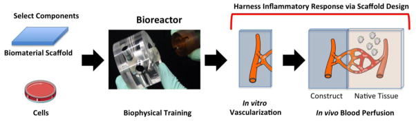
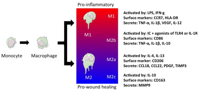
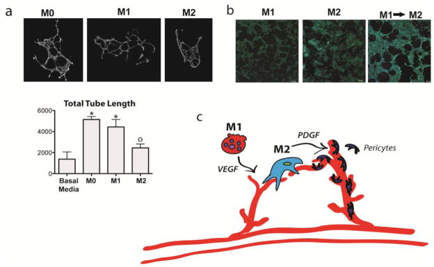
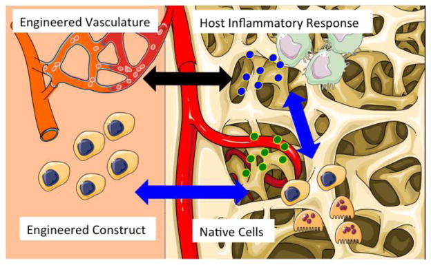
Tissue engineering is increasingly based on recapitulating human physiology, through integration of biological principles into engineering designs. In spite of all progress in engineering functional human tissues, we are just beginning to develop effective methods for establishing blood perfusion and controlling the inflammatory factors following implantation into the host. Functional vasculature largely determines tissue survival and function in vivo. The inflammatory response is a major regulator of vascularization and overall functionality of engineered tissues, through the activity of different types of macrophages and the cytokines they secrete. We discuss here the cell-scaffold-bioreactor systems for harnessing the inflammatory response for enhanced tissue vascularization and healing. To this end, inert scaffolds that have been considered for many decades a "gold standard" in regenerative medicine are beginning to be replaced by a new generation of "smart" tissue engineering systems designed to actively mediate tissue survival and function.


Figures

Figure 1
Figure 1. Biomaterial scaffold
Schematic showing the steps towards an engineered construct where the scaffold is designed to harness the host tissue response and maximize the angiogenic response. The first step is to select the cell source and the biomaterial of interest. These two components are then assembled and trained in a bioreactor by applying tissue appropriate biophysical forces. At this stage, a vasculature can be formed and perfused in vitro. This “artificial vasculature” can then be connected to the host tissue vasculature by harnessing the inflammatory response at the site of implantation. Artwork provided by Servier Medical Arts.
Figure 2
Figure 2. Differentiation and polarization of macrophages
Monocytes that are recruited to the site of injury differentiate into macrophages via local signals. These macrophages polarize into a spectrum of macrophage subtypes ranging from pro-inflammatory (M1) to wound healing phenotypes (M2c). Artwork provided by Servier Medical Arts.
Figure 3
Figure 3. Contributions of M1 and M2 macrophages to angiogenesis
(a) M1 but not M2 macrophages increase sprouting of endothelial cells on Matrigel (* indicates p<0.05 and o indicates p>0.05 compared to control, one-way ANOVA and Tukey’s post-hoc analysis). Networks were stained with Live/Dead kit (Invitrogen), imaged at 10x magnification with an Olympus IX81 fluorescent microscope, and analyzed with ImageJ’s Angiogenesis Analyzer macro. (b) Endothelial cells organized into a loosely connected network on fibrin gel when the media is changed from M1-conditioned media to M2-conditioned media, which did not occur when endothelial cells were cultured uniformly in either M1- or M2-conditioned media. Endothelial cells were stained with DAPI and phalloidin tagged with Alexafluor-488 and imaged at 10x with an Olympus Fluoview FV1000 confocal microscope. (c) Proposed model of macrophage control over angiogenesis, in which M1 macrophages initiate sprouting via VEGF and M2 macrophages promote vessel stabilization via guiding anastomosis and possibly recruiting pericytes via PDGF. (Figure modified with permission from ). Artwork provided by Servier Medical Arts.
Figure 4
Figure 4. Immunomodulation of engineered tissues for enhanced vascularization and healing
Advanced tissue engineering strategies will take into account interactions between the cells and vasculature of engineered tissue with the native cells and inflammatory response of the host environment. Artwork provided by Servier Medical Arts.

References

    1. Agrawal H, Tholpady SS, Capito AE, Drake DB, Katz AJ. Macrophage phenotypes correspond with remodeling outcomes of various acellular dermal matrices. Open Journal of Regenerative Medicine. 2012;1:51–59.
    1. Alexander KA, Chang MK, Maylin ER, Kohler T, Muller R, Wu AC, Van Rooijen N, Sweet MJ, Hume DA, Raggatt LJ, Pettit AR. Osteal macrophages promote in vivo intramembranous bone healing in a mouse tibial injury model. J Bone Miner Res. 2011;26:1517–1532. - PubMed
    1. Anghelina M, Krishnan P, Moldovan L, Moldovan NI. Monocytes and macrophages form branched cell columns in matrigel: implications for a role in neovascularization. Stem Cells Dev. 2004;13:665–676. - PubMed
    1. Anghelina M, Krishnan P, Moldovan L, Moldovan NI. Monocytes/macrophages cooperate with progenitor cells during neovascularization and tissue repair: conversion of cell columns into fibrovascular bundles. Am J Pathol. 2006;168:529–541. - PMC - PubMed
    1. Arendt LM, McCready J, Keller PJ, Baker DD, Naber SP, Seewaldt V, Kuperwasser C. Obesity Promotes Breast Cancer by CCL2-Mediated Macrophage Recruitment and Angiogenesis. Cancer Res. 2013;73:6080–6093. - PMC - PubMed

Publication types

LinkOut - more resources