. 2015 Jan 1;197(1):128-37.
doi: 10.1128/JB.02063-14. Epub 2014 Oct 20.

RelA inhibits Bacillus subtilis motility and chaining

Affiliations

RelA inhibits Bacillus subtilis motility and chaining

Qutaiba O Ababneh et al. J Bacteriol. .

Abstract

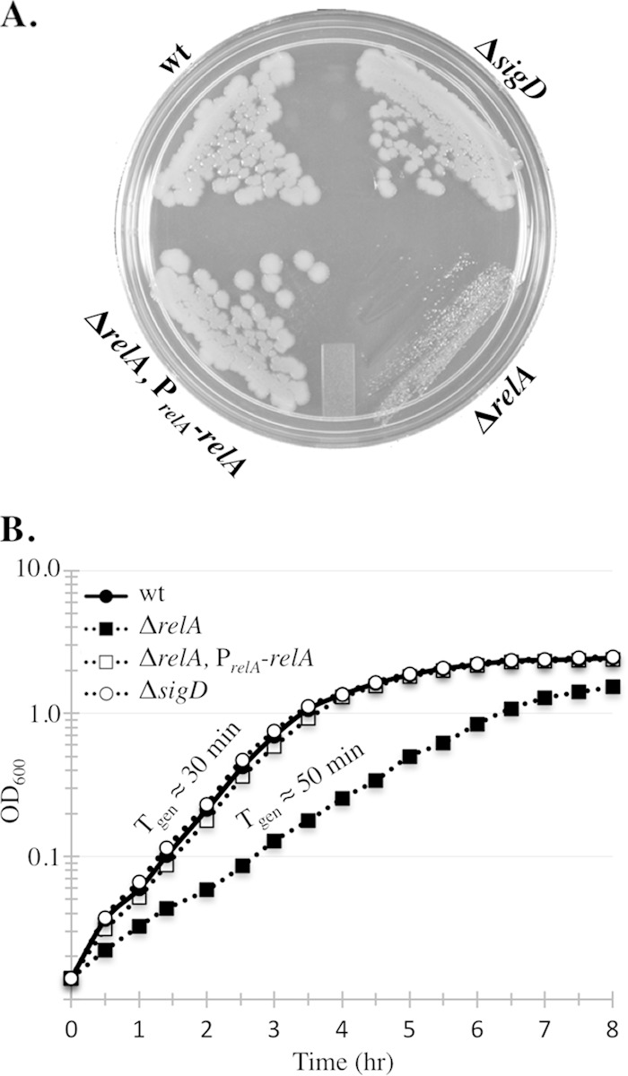
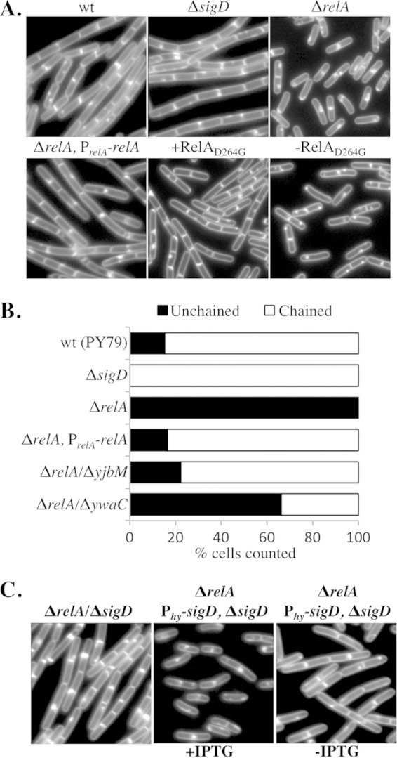
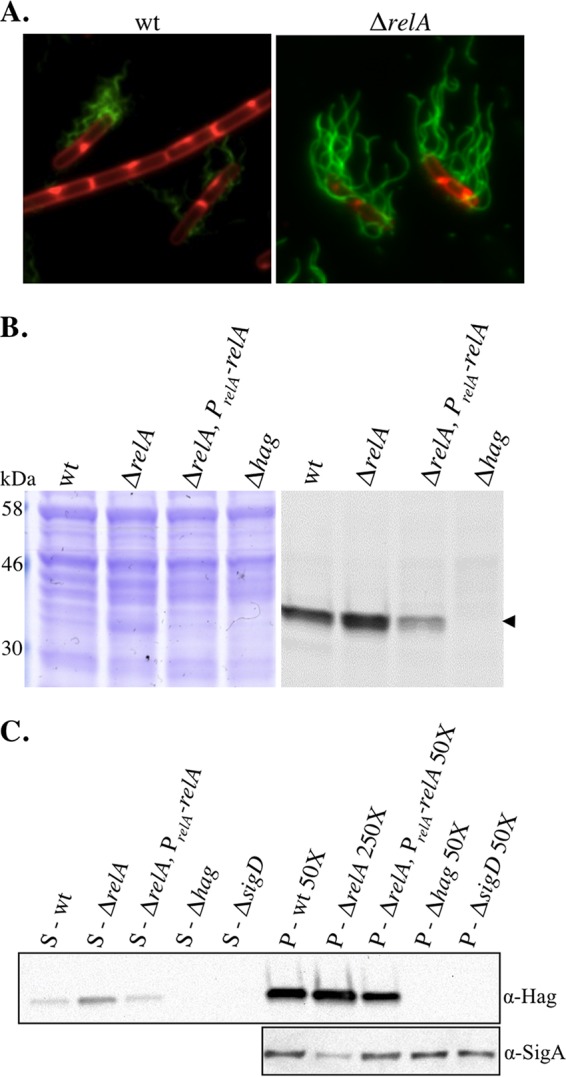
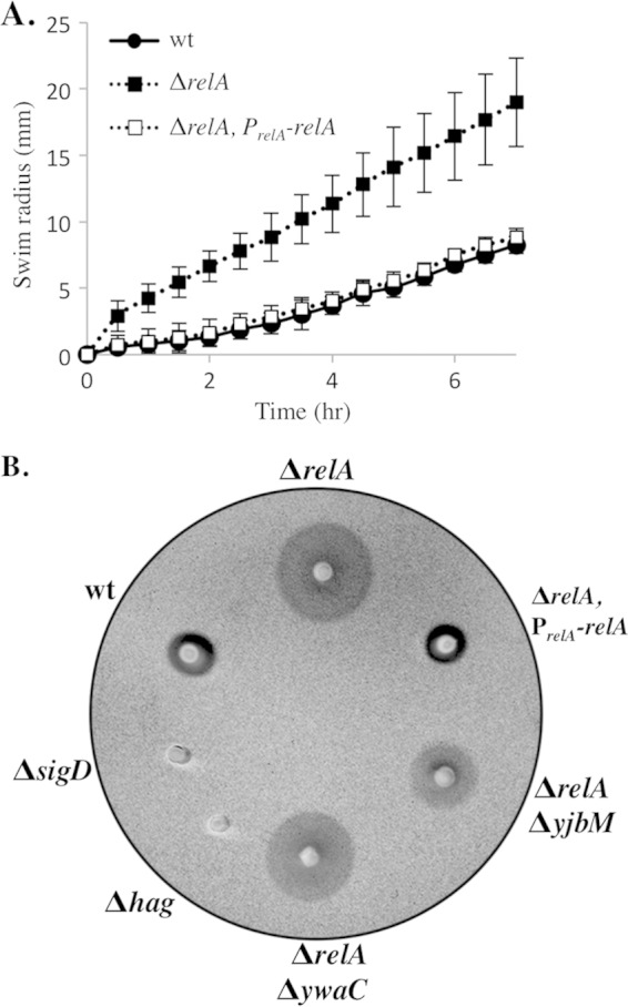
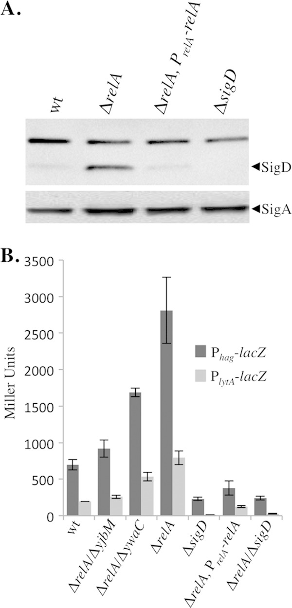
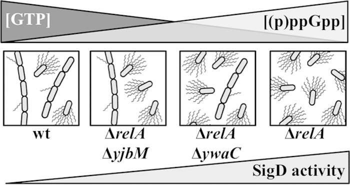
The nucleotide second messengers pppGpp and ppGpp [(p)ppGpp] are responsible for the global downregulation of transcription, translation, DNA replication, and growth rate that occurs during the stringent response. More recent studies suggest that (p)ppGpp is also an important effector in many nonstringent processes, including virulence, persister cell formation, and biofilm production. In Bacillus subtilis, (p)ppGpp production is primarily determined by the net activity of RelA, a bifunctional (p)ppGpp synthetase/hydrolase, and two monofunctional (p)ppGpp synthetases, YwaC and YjbM. We observe that in B. subtilis, a relA mutant grows exclusively as unchained, motile cells, phenotypes regulated by the alternative sigma factor SigD. Our data indicate that the relA mutant is trapped in a SigD "on" state during exponential growth, implicating RelA and (p)ppGpp levels in the regulation of cell chaining and motility in B. subtilis. Our results also suggest that minor variations in basal (p)ppGpp levels can significantly skew developmental decision-making outcomes.


Figures

FIG 1
FIG 1
The relA mutant grows slowly and exponentially. (A) The indicated strains were streaked on an LB plate and incubated for 24 h at 37°C. (B) Growth curves of the same strains shown panel A grown in liquid CH medium. The average of three independent replicates is plotted for each strain. wt, wild type (BJH001); ΔrelA, BQA009; ΔrelA PrelA-relA, BQA068; ΔsigD, BQA022.
FIG 2
FIG 2
The ΔrelA mutant grows exclusively as single cells. (A) Representative images of wt (BJH001), ΔrelA (BQA009), ΔrelA PrelA-relA (BQA068), ΔsigD (BQA022), and Pspac-relAD264G (EUB004) cells grown to mid-log phase in CH medium. For EUB004, cells grown in the presence (+RelAD264G) and absence (−RelAD264G) of 1 mM IPTG for 120 min are shown. The membranes were stained with TMA. (B) Frequencies of chained (three or more cells linked) and unchained (single and doublet) cells across a population. The cells in images from at least three independent cultures were counted, and no fewer than 1,500 cells were quantitated for each strain represented in the graph. (C) Representative images of ΔrelA ΔsigD (BQA059) and ΔrelA Phy-sigD ΔsigD (BQA083) cells grown to exponential phase in CH medium. The membranes were stained with TMA.
FIG 3
FIG 3
The relA mutant is comprised of mostly flagellated cells (A) Representative images of mid-log-phase cultures of wt (BQA057) and ΔrelA (BQA062) strains. The membranes were stained with FM4-64 (red), and flagellin (Hag) was stained with Alexa Fluor 488 C5 maleimide (green). Both strains harbor a point mutation in the chromosomal copy of hag that creates HagT209C. (B) Comparison of flagellin (Hag) protein levels in wt (BQA057), ΔrelA (BQA062), ΔrelA PrelA-relA (BQA080), and Δhag (BQA076) strains. Samples were collected from mid-log-phase cultures grown in CH medium and stained with Alexa Fluor 488 C5 maleimide. Proteins from cell lysates were separated by SDS-PAGE, stained with Coomassie blue (left), and scanned with a laser scanner to visualize fluorescently labeled protein (right). The arrowhead indicates flagellin. (C) (Top) Western blot analysis showing flagellin levels associated with the culture supernatants (left; S) and cell pellets (right; P) of the indicated strains grown in CH medium to mid-log phase: wt (BJH001), ΔrelA (BQA009), ΔrelA PrelA-relA (BQA068), ΔsigD (BQA022), and Δhag (BQA076). It was necessary to dilute the cell pellet lysates to the indicated dilutions in order to assess the levels of protein associated with each sample without overexposure. (Bottom) SigA protein served as a loading control for the cell pellet samples.
FIG 4
FIG 4
Loss of relA leads to increased mobility on swim plates. (A) Quantitation of swim expansion from plate assays performed with wt (BJH001), ΔrelA (BQA009), and ΔrelA PrelA-relA (BQA068) strains on LB fortified with 0.25% agar. Each symbol indicates the average of measurements from five independent experiments with standard deviations. Measurements were recorded every 30 min for 8 h. (B) wt (BJH001), ΔrelA (BQA009), ΔrelA PrelA-relA (BQA068), ΔsigD (BQA022), Δhag (BQA076), ΔrelA ΔyjbM (BQA081), and ΔrelA ΔywaC (BQA082) strains were inoculated on LB fortified with 0.25% agar. After 3.5 h of incubation at 37°C, the plates were scanned against a black background. The images were inverted to better show contrast.
FIG 5
FIG 5
Loss of relA leads to an increase in SigD levels and activity. (A) Comparison of SigD protein levels in wt (BJH001), ΔrelA (BQA009), ΔrelA PrelA-relA (BQA068), and ΔsigD (BQA022) strains. Samples were collected from mid-log-phase cultures grown in CH medium. Proteins from cell lysates were separated by SDS-PAGE and analyzed by Western blot analysis by probing with either anti-SigD or anti-SigA antibody, as indicated. (B) β-Galactosidase assays of Phag-lacZ and PlytA-lacZ transcriptional activities conducted on wt (BJH046 and BJH047), ΔrelA (BQA050 and BQA051), ΔrelA ΔyibM (BQA086 and BQA087), ΔrelA ΔywaC (BQA088 and BQA089), ΔsigD (BQA071 and BQA072), ΔrelA PrelA-relA (BQA073 and BQA074), and ΔrelA ΔsigD (BQA084 and BQA085) strains. Samples were collected from mid-log-phase cultures grown in CH medium. The data shown are the means of three independent replicates with standard deviations.
FIG 6
FIG 6
Model depicting the observed relationships between cell motility and chaining, SigD activity, GTP concentration (22), and putative (p)ppGpp levels in various (p)ppGpp synthetase/hydrolase backgrounds.

References

    1. Dubnau D, Losick R. 2006. Bistability in bacteria. Mol Microbiol 61:564–572. doi:10.1111/j.1365-2958.2006.05249.x. - DOI - PubMed
    1. Shank EA, Kolter R. 2011. Extracellular signaling and multicellularity in Bacillus subtilis. Curr Opin Microbiol 14:741–747. doi:10.1016/j.mib.2011.09.016. - DOI - PMC - PubMed
    1. Mirouze N, Desai Y, Raj A, Dubnau D. 2012. Spo0A∼P imposes a temporal gate for the bimodal expression of competence in Bacillus subtilis. PLoS Genet 8:e1002586. doi:10.1371/journal.pgen.1002586. - DOI - PMC - PubMed
    1. Cozy LM, Phillips AM, Calvo RA, Bate AR, Hsueh YH, Bonneau R, Eichenberger P, Kearns DB. 2012. SlrA/SinR/SlrR inhibits motility gene expression upstream of a hypersensitive and hysteretic switch at the level of sigma(D) in Bacillus subtilis. Mol Microbiol 83:1210–1228. doi:10.1111/j.1365-2958.2012.08003.x. - DOI - PMC - PubMed
    1. Kearns DB, Losick R. 2005. Cell population heterogeneity during growth of Bacillus subtilis. Genes Dev 19:3083–3094. doi:10.1101/gad.1373905. - DOI - PMC - PubMed

Publication types

MeSH terms

LinkOut - more resources