. 2015 Jan 1;197(1):201-10.
doi: 10.1128/JB.02015-14. Epub 2014 Oct 20.

The rv1184c locus encodes Chp2, an acyltransferase in Mycobacterium tuberculosis polyacyltrehalose lipid biosynthesis

Affiliations

The rv1184c locus encodes Chp2, an acyltransferase in Mycobacterium tuberculosis polyacyltrehalose lipid biosynthesis

Megan H Touchette et al. J Bacteriol. .

Abstract

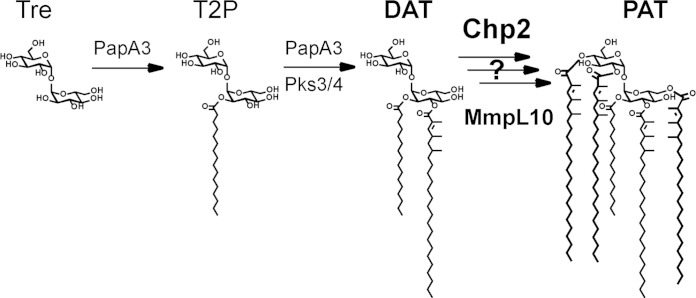
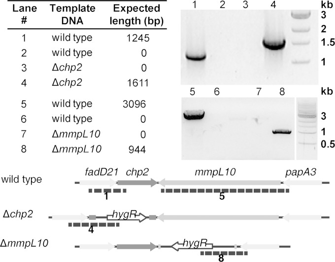
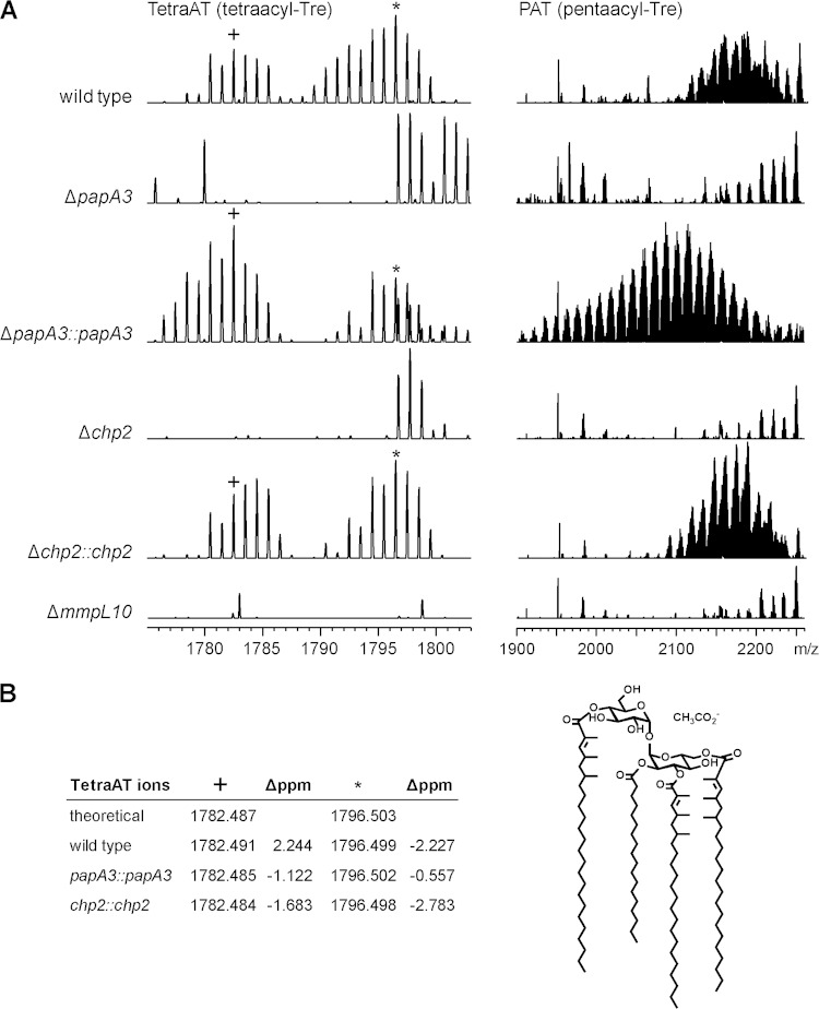
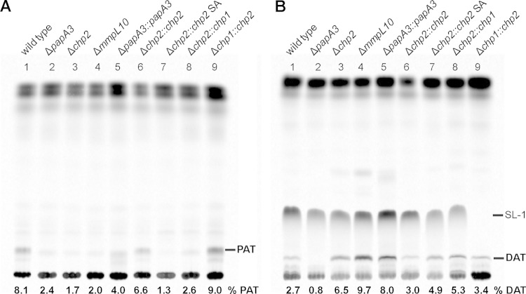
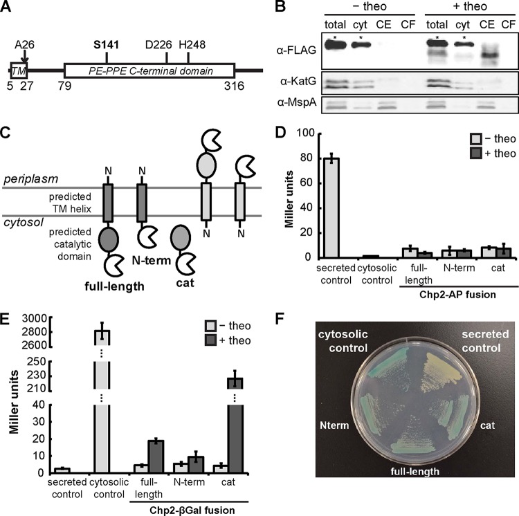
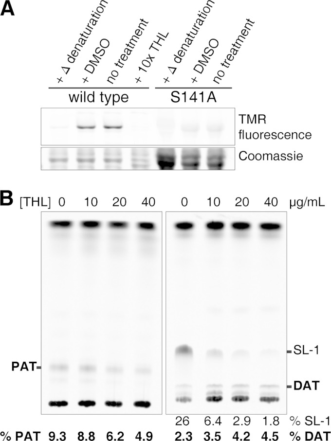
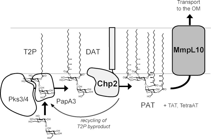
Trehalose glycolipids are found in many bacteria in the suborder Corynebacterineae, but methyl-branched acyltrehaloses are exclusive to virulent species such as the human pathogen Mycobacterium tuberculosis. In M. tuberculosis, the acyltransferase PapA3 catalyzes the formation of diacyltrehalose (DAT), but the enzymes responsible for downstream reactions leading to the final product, polyacyltrehalose (PAT), have not been identified. The PAT biosynthetic gene locus is similar to that of another trehalose glycolipid, sulfolipid 1. Recently, Chp1 was characterized as the terminal acyltransferase in sulfolipid 1 biosynthesis. Here we provide evidence that the homologue Chp2 (Rv1184c) is essential for the final steps of PAT biosynthesis. Disruption of chp2 led to the loss of PAT and a novel tetraacyltrehalose species, TetraAT, as well as the accumulation of DAT, implicating Chp2 as an acyltransferase downstream of PapA3. Disruption of the putative lipid transporter MmpL10 resulted in a similar phenotype. Chp2 activity thus appears to be regulated by MmpL10 in a relationship similar to that between Chp1 and MmpL8 in sulfolipid 1 biosynthesis. Chp2 is localized to the cell envelope fraction, consistent with its role in DAT modification and possible regulatory interactions with MmpL10. Labeling of purified Chp2 by an activity-based probe was dependent on the presence of the predicted catalytic residue Ser141 and was inhibited by the lipase inhibitor tetrahydrolipstatin (THL). THL treatment of M. tuberculosis resulted in selective inhibition of Chp2 over PapA3, confirming Chp2 as a member of the serine hydrolase superfamily. Efforts to produce in vitro reconstitution of acyltransferase activity using straight-chain analogues were unsuccessful, suggesting that Chp2 has specificity for native methyl-branched substrates.


Figures

FIG 1
FIG 1
The biosynthetic pathway for PAT is incomplete. The acyltransferase PapA3 converts trehalose into T2P and then to DAT using mycolipenic chains produced by Pks3/4. Chp2 and MmpL10 may be involved in the subsequent conversion of DAT into PAT.
FIG 2
FIG 2
Confirmation of strains Δchp2 and ΔmmpL10 by PCR. For chp2, the 5′ primer annealed ∼900 bp upstream of chp2. For lanes 1 and 3, the 3′ primer annealed to a region of chp2 that is absent in the knockout strain. For mmpL10, the 5′ primer annealed ∼100 bp upstream of mmpL10. For lanes 5 and 7, the 3′ primer annealed to a region of mmpL10 that is absent in the knockout strain. For lanes 2, 4, 6, and 8, the 3′ primer annealed to the hygR marker that replaces chp2 and mmpL10 in the knockout strains. See Table S2 in the supplemental material for oligonucleotide sequences.
FIG 3
FIG 3
Chp2 and MmpL10 are required for biosynthesis of PAT and the novel acyltrehalose TetraAT. M. tuberculosis wild-type, ΔpapA3, Δchp2, and ΔmmpL10 strains were analyzed by ESI-MS. (A) TetraAT and PAT were absent from all three mutants but were restored in the ΔpapA3 and Δchp2 complement strains. PAT appears as a characteristic envelope of peaks centered at approximately m/z 2,100 to 2,200. A representative segment of the TetraAT spectrum is shown in order to highlight two of the peaks used for assignment. (B) TetraAT was assigned by exact mass, and observed ions were within 3 ppm of the predicted m/z value. As an example, a possible structure for the m/z 1,782.487 [M+CH3CO2-] ion is shown.
FIG 4
FIG 4
Chp2 and MmpL10 function downstream of DAT, and Chp2 is specific to the PAT biosynthesis. M. tuberculosis wild-type, ΔpapA3, Δchp2, and ΔmmpL10 strains were analyzed by metabolic labeling with [14C]propionate followed by TLC analysis of lipid extracts. In addition to loss of PAT in all mutants (lanes 2 to 4) (A), DAT was absent from the ΔpapA3 mutant (lane 2) (B). PAT and DAT were restored upon complementation (lanes 5 and 6), except in the Δchp2::chp2 SA mutant (lane 7). The chp1 and chp2 homologues did not cross-complement the Δchp2 and Δchp1 mutants (lanes 8 and 9). The mobile phase was 92:8 petroleum ether/acetone (A) and 90:10 chloroform/methanol (B) (21, 29, 30). The proportion of PAT or DAT is reported as the percent total integrated intensity in each lane.
FIG 5
FIG 5
Chp2 localizes to the cell envelope with the catalytic domain in the cytosol. (A) Primary structure of Chp2 with the predicted positions of the transmembrane helix (TM), signal peptide cleavage site (A27), conserved domain, and catalytic residues. (B) Immunoblot of subcellular fractions of M. smegmatis expressing theophylline (theo)-inducible Chp2-3xFLAG showed Chp2 enrichment in the cell envelope (CE) fraction. KatG and MspA are markers for the cytosol (cyt) and outer membrane, respectively. Each asterisk indicates a nonspecific α-FLAG-reactive band. (C) AP or βGal was expressed in M. smegmatis as a theophylline-inducible fusion(s) to the C terminus of full-length Chp2, the N-terminal domain, or the catalytic domain. (D to F) Enzymatic activity was not detected for the AP fusions (D) but was observed for the βGal fusions (E), which were further confirmed by growth on chromogenic medium containing X-Gal (F). Strains grown without theophylline or expressing AP or βGal with or without the Ag85B secretion signal served as negative and positive controls (48). Turnover of colorimetric substrates is expressed in Miller units.
FIG 6
FIG 6
Chp2 is a serine hydrolase-type enzyme that is inhibited by THL. (A) Labeling of purified Chp2-cat domain by TMR-FP was detected by fluorescence and inhibited by heat denaturation, treatment with THL, or mutation of the catalytic residue (S141A). (B) Lipid extracts from M. tuberculosis treated with different concentrations of THL for 6 h followed by [14C]propionate labeling revealed the dose-dependent, but incomplete, inhibition of PAT and accumulation of DAT (mobile phases were as described in the Fig. 4 legend). SL-1 inhibition by THL has been previously reported (18). The proportion of PAT or DAT is reported as percent total integrated intensity in each lane (SL-1 results are shown for comparison).
FIG 7
FIG 7
Proposed model for acyltrehalose biosynthesis and transmembrane transport. The data are most consistent with a model in which Chp2 completes PAT biosynthesis in the cytosolic leaflet and MmpL10 transports acyltrehaloses across the membrane. The AcTre side products generated by Chp2 can be recycled by PapA3. (Note that the mycolipenic groups are truncated in this figure for simplicity.) OM, outer membrane.

References

    1. Khan AA, Stocker BL, Timmer MS. 2012. Trehalose glycolipids–synthesis and biological activities. Carbohydr Res 356:25–36. doi: 10.1016/j.carres.2012.03.010. - DOI - PubMed
    1. Daffé M, Lacave C, Lanéelle MA, Gillois M, Lanéelle G. 1988. Polyphthienoyl trehalose, glycolipids specific for virulent strains of the tubercle bacillus. Eur J Biochem 172:579–584. doi: 10.1111/j.1432-1033.1988.tb13928.x. - DOI - PubMed
    1. Ortalo-Magné A, Lemassu A, Lanéelle MA, Bardou F, Silve G, Gounon P, Marchal G, Daffé M. 1996. Identification of the surface-exposed lipids on the cell envelopes of Mycobacterium tuberculosis and other mycobacterial species. J Bacteriol 178:456–461. - PMC - PubMed
    1. Dubey VS, Sirakova TD, Kolattukudy PE. 2002. Disruption of msl3 abolishes the synthesis of mycolipanoic and mycolipenic acids required for polyacyltrehalose synthesis in Mycobacterium tuberculosis H37Rv and causes cell aggregation. Mol Microbiol 45:1451–1459. doi: 10.1046/j.1365-2958.2002.03119.x. - DOI - PubMed
    1. Rousseau C, Neyrolles O, Bordat Y, Giroux S, Sirakova TD, Prevost M-C, Kolattukudy PE, Gicquel B, Jackson M. 2003. Deficiency in mycolipenate- and mycosanoate-derived acyltrehaloses enhances early interactions of Mycobacterium tuberculosis with host cells. Cell Microbiol 5:405–415. doi: 10.1046/j.1462-5822.2003.00289.x. - DOI - PubMed

Publication types

MeSH terms

Associated data