. 2015 Jan;35(1):76-87.
doi: 10.1128/MCB.00764-14. Epub 2014 Oct 20.

Axl phosphorylates Elmo scaffold proteins to promote Rac activation and cell invasion

Affiliations

Axl phosphorylates Elmo scaffold proteins to promote Rac activation and cell invasion

Afnan Abu-Thuraia et al. Mol Cell Biol. 2015 Jan.

Abstract

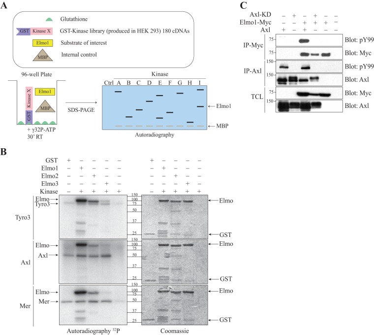
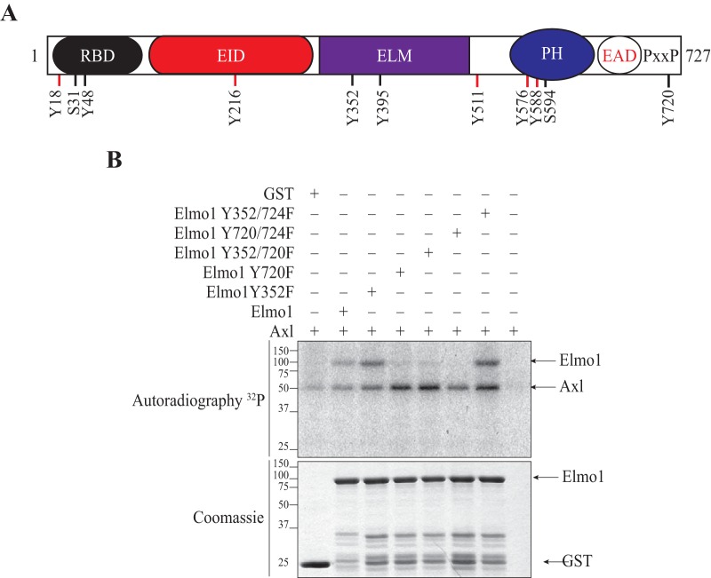
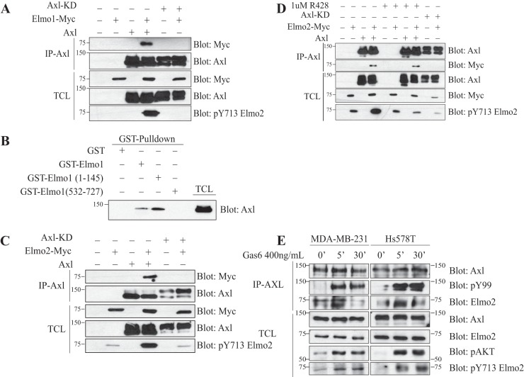
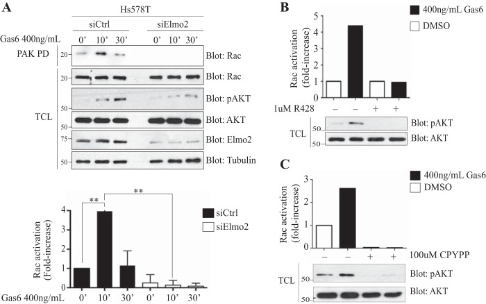
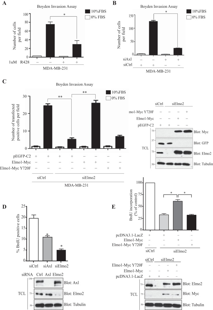
The receptor tyrosine kinase Axl contributes to cell migration and invasion. Expression of Axl correlates with metastatic progression in cancer patients, yet the specific signaling events promoting invasion downstream of Axl are poorly defined. Herein, we report Elmo scaffolds to be direct substrates and binding partners of Axl. Elmo proteins are established to interact with Dock family guanine nucleotide exchange factors to control Rac-mediated cytoskeletal dynamics. Proteomics and mutagenesis studies reveal that Axl phosphorylates Elmo1/2 on a conserved carboxyl-terminal tyrosine residue. Upon Gas6-dependent activation of Axl, endogenous Elmo2 becomes phosphorylated on Tyr-713 and enters into a physical complex with Axl in breast cancer cells. Interfering with Elmo2 expression prevented Gas6-induced Rac1 activation in breast cancer cells. Similarly to blocking of Axl, Elmo2 knockdown or pharmacological inhibition of Dock1 abolishes breast cancer cell invasion. Interestingly, Axl or Elmo2 knockdown diminishes breast cancer cell proliferation. Rescue of Elmo2 knockdown cells with the wild-type protein but not with Elmo2 harboring Tyr-713-Phe mutations restores cell invasion and cell proliferation. These results define a new mechanism by which Axl promotes cell proliferation and invasion and identifies inhibition of the Elmo-Dock pathway as a potential therapeutic target to stop Axl-induced metastases.


Figures

FIG 1
FIG 1
The TAM receptors phosphorylate Elmo proteins. (A) Schematic overview of the kinase screen. Elmo1 is the substrate of interest, and MBP1 is used as a positive control. (B) Elmo is phosphorylated in vitro by the TAMs. An in vitro kinase assay was performed, where 2 μg of GST-Elmo proteins were incubated with 0.05 μg of the kinase domains of Tyro3, Axl and Mer, and [γ-32P]ATP. Expression of the proteins was analyzed by Coomassie staining and the phosphorylation by autoradiography. (C) Elmo1 phosphorylation in cells is dependent on Axl catalytic activity. Lysates of HEK293T cells transfected with the indicated plasmids were immunoprecipitated with an antibody against the Myc epitope (Elmo1) and with an antibody against Axl. The phosphorylation and expression levels of Elmo1 and Axl were analyzed via immunoblotting with anti-Myc and anti-Axl antibodies, respectively.
FIG 2
FIG 2
Elmo1 is phosphorylated on tyrosines 720 and 724 by TAM receptors. (A) Elmo1 phosphorylation sites identified by mass spectrometry. Lysates of HEK293T cells were transfected with c-Myc–Elmo1 and were either subjected to an in vitro kinase assay with GST-Tyro3 or cotransfected with GST-Tyro3 and subjected to immunoprecipitation of c-Myc–Elmo1. Sites on Elmo1 phosphorylated by Tyro3 were identified by mass spectrometry. Sites depicted in red were identified by both experiments, whereas the sites depicted in black were identified only in the c-Myc–Elmo1 immunoprecipitation. RBD, Ras-binding domain; EID, Elmo inhibitory domain; ELM, Elmo domain; PH, atypical pleckstrin homology domain; EAD, Elmo autoregulatory domain; PxxP, proline-rich region. (B) Elmo1 is phosphorylated at tyrosines 720 and 724. An in vitro kinase assay was performed, where 3 μg of the GST-Elmo1 protein or Y-F mutants were incubated with 0.025 μg of the Axl kinase domain and [γ-32P]ATP. Expression of the proteins were analyzed by Coomassie staining and phosphorylation by autoradiography.
FIG 3
FIG 3
Elmo modulation by Axl is dependent on Axl's catalytic activity. (A and C) Axl that is wild type and not kinase dead interacts with and phosphorylates Elmo1 (A) and Elmo2 (C). Lysates of HEK293T cells transfected with the indicated plasmids were coimmunoprecipitated with an antibody against Axl. The coprecipitation and expression levels of the Axl proteins and Elmo1 (A) or Elmo2 (C) were analyzed via immunoblotting with anti-Myc (Elmo) and anti-Axl antibodies. (B) Interaction between Elmo1 and Axl is mediated by the N terminus of Elmo1. Lysates from transfected HEK293T cells with Axl were incubated with 3 μg of the GST-Elmo1 full-length protein or fragments. The binding of Axl to Elmo1 fragments and the expression levels were analyzed via immunoblotting with anti-Axl antibody. (D) Inhibition of Axl activity with R428 abolishes Elmo2 phosphorylation by Axl. Transfected HEK293T cells with the indicated plasmids were serum starved prior to treatment with either dimethyl sulfoxide (DMSO) or a 1 μM concentration of the Axl inhibitor R428 for 1 h. Lysates were coimmunoprecipitated with an antibody against Axl. The coprecipitation and expression levels of the Axl proteins and Elmo2 was analyzed via immunoblotting with anti-Myc (Elmo2) and anti-Axl antibodies, respectively. Protein expression and Elmo2 phosphorylation were analyzed by immunoblotting with anti-Myc, anti-Axl, and anti-pY713 Elmo2. (E) Axl interacts with and phosphorylates endogenous Elmo2 in basal breast cancer cell lines. MDA-MB-231 and Hs578T cells were treated with 400 ng/ml of Gas6 for the indicated time points and then were subjected to coimmunoprecipitation of Axl using anti-Axl antibody. The precipitation of Elmo2 by Axl was detected by immunoblotting with anti-Elmo2 antibody. The protein expression levels were also analyzed via immunoblotting with anti-pAKT, anti-pY99, anti-Elmo2, anti-Axl, and anti-pY713 Elmo2 antibodies.
FIG 4
FIG 4
Rac activation in Hs578T cells is Axl and Elmo2 dependent. (A) Elmo2 is required for Rac activation upon Gas6 stimulation. Hs578T cells transfected with 60 nM NON-Targeting or ON-Target SmartPool Elmo2 siRNA prior to being treated with 400 ng/ml of Gas6 at the indicated time points were assayed for Rac activation by precipitation of Rac with the purified p21-binding domain of PAK protein kinase expressed as a GST fusion protein (GST-PAK-PBD) (n = 6). The amount of Rac in pulldowns and in total cell lysates (TCL) was detected by immunoblotting with an anti-Rac antibody. Expression levels of the various proteins and equal loading of Rac in all samples were analyzed by immunoblotting of the TCL using anti-Rac, antitubulin, anti-Elmo2, anti-pAKT, and anti-AKT. (B and C) Axl and Dock1 inhibition reduces Rac activation upon Gas6 stimulation. Hs578T cells were treated with 1 μM R428 (B) or with 100 μM CPYPP (C) for 1 h followed by 400 ng/ml Gas6 for 20 min. Rac activation was assayed by precipitation with the purified p21-binding domain of PAK protein kinase expressed as a GST fusion protein (GST-PAK-PBD) (n = 5). The amount of Rac in pulldowns and in total cell lysates (TCL) was quantified by the software program Image J. Expression levels of the various proteins were analyzed by immunoblotting of the TCL using anti-pAKT and anti-AKT. Data are shown as means ± SD; **, P < 0.0001; one-way ANOVA.
FIG 5
FIG 5
Cell invasion and proliferation of MDA-MB-231 cells is Elmo2 and Axl dependent. (A and B) Axl activity or expression inhibition reduces cell invasion. Serum-starved MDA-MB-231 cells treated with 1 μM R428 (A) or transfected with 100 nM NON-Targeting or ON-Target SmartPool Axl siRNA (B) were detached and placed in the upper compartment of a Boyden chamber. Cells were allowed to invade through the Matrigel for 16 h. The invasion assay was performed in triplicate, and data are shown as means ± SEM; *, P < 0.001; one-way ANOVA. (C) The Elmo1 phosphorylation site Tyr 720 is required for cell invasion in MDA-MB-231 cells. Serum-starved MDA-MB-231 cells transfected with 60 nM NON-Targeting or ON-Target SmartPool Elmo2 siRNA and rescued 24 h later with either 1 μg GFP or 1 μg Elmo1 wild type or 2 μg Elmo1 Y720F mutant were detached and placed in the upper compartment of a Boyden chamber. Cells were allowed to invade through the Matrigel for 16 h and then were fixed and stained with anti-Myc and anti-GFP. GFP- and Myc-positive cells that invaded the Matrigel to the underside of the membrane were counted from photographs taken at magnification ×20. The invasion assay was performed in triplicate, and data are shown as mean ± SD; **, P < 0.0001; one-way ANOVA. Expression levels of the transfected proteins for invasion assays were analyzed by immunoblotting cell lysates with anti-Elmo2, anti-Myc, anti-GFP, and antitubulin antibodies, as indicated. (D and E) Axl and Elmo1 phosphorylation site Tyr 720 are required for cell proliferation in MDA-MB-231 cells. Cells transfected with 100 nM NON-Targeting, 100 nM ON-Target SmartPool Axl siRNA, or 60 nM ON-Target SmartPool Elmo2 siRNA were detached 24 h later and plated on fibronectin-coated glass slips. Transfected MDA-MB-231 cells with 60 nM NON-Targeting or ON-Target SmartPool Elmo2 siRNA were rescued 24 h later with either 1 μg pcDNA3.1-LacZ or 1 μg Elmo1 wild type or 2 μg Elmo1 Y720F mutant prior to being plated on fibronectin-coated glass slips 24 h later (E). Cells were allowed to grow for 24 h and then were stained with BrdU for 30 min. The BrdU-positive cells were counted, and the percentage of BrdU-positive cells versus total cells stained for DAPI were calculated in 5 different fields of each condition. Three experiments were performed, and the percentage of BrdU-positive cells was calculated for each experiment and used for the final quantification. For rescue experiments (E), BrdU incorporation was expressed as a percentage of decrease relative to control (n = 3). Values are reported as means ± SEM. Expression levels of the transfected proteins for proliferation assays were analyzed by immunoblotting cell lysates with anti-Elmo2, anti-Myc, anti-Axl, and antitubulin antibodies, as indicated.

References

    1. O'Bryan JP, Frye RA, Cogswell PC, Neubauer A, Kitch B, Prokop C, Espinosa R III, Le Beau MM, Earp HS, Liu ET. 1991. axl, a transforming gene isolated from primary human myeloid leukemia cells, encodes a novel receptor tyrosine kinase. Mol Cell Biol 11:5016–5031. - PMC - PubMed
    1. Linger RM, Keating AK, Earp HS, Graham DK. 2008. TAM receptor tyrosine kinases: biologic functions, signaling, and potential therapeutic targeting in human cancer. Adv Cancer Res 100:35–83. doi:10.1016/S0065-230X(08)00002-X. - DOI - PMC - PubMed
    1. Stitt TN, Conn G, Gore M, Lai C, Bruno J, Radziejewski C, Mattsson K, Fisher J, Gies DR, Jones PF, Masiakowski P, Ryan TE, Tobkes NJ, Chen DH, DiStefano PS, Long GL, Basilico C, Goldfarb MP, Lemke G, Glass DJ, Yancopoulos GD. 1995. The anticoagulation factor protein S and its relative, Gas6, are ligands for the Tyro 3/Axl family of receptor tyrosine kinases. Cell 80:661–670. doi:10.1016/0092-8674(95)90520-0. - DOI - PubMed
    1. Caberoy NB, Zhou Y, Li W. 2010. Tubby and tubby-like protein 1 are new MerTK ligands for phagocytosis. EMBO J 29:3898–3910. doi:10.1038/emboj.2010.265. - DOI - PMC - PubMed
    1. Nagata K, Ohashi K, Nakano T, Arita H, Zong C, Hanafusa H, Mizuno K. 1996. Identification of the product of growth arrest-specific gene 6 as a common ligand for Axl, Sky, and Mer receptor tyrosine kinases. J Biol Chem 271:30022–30027. doi:10.1074/jbc.271.47.30022. - DOI - PubMed

Publication types

MeSH terms