. 2015 Jan;161(Pt 1):18-29.
doi: 10.1099/mic.0.083378-0. Epub 2014 Oct 20.

Functional regions of Candida albicans hyphal cell wall protein Als3 that determine interaction with the oral bacterium Streptococcus gordonii

Affiliations

Functional regions of Candida albicans hyphal cell wall protein Als3 that determine interaction with the oral bacterium Streptococcus gordonii

Caroline V Bamford et al. Microbiology (Reading). 2015 Jan.

Abstract

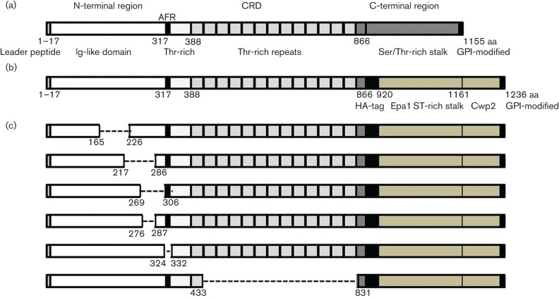
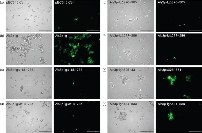
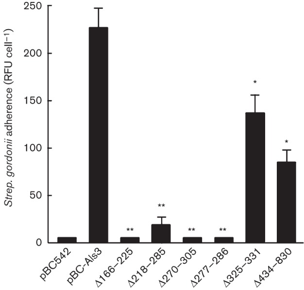
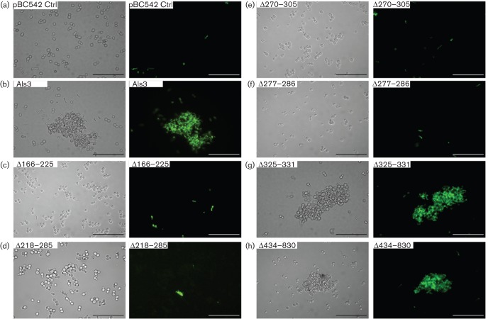
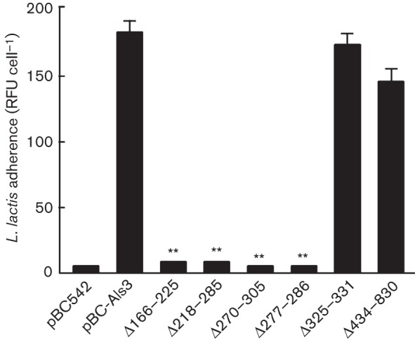
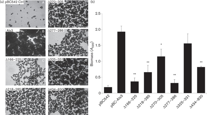
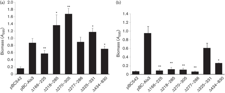
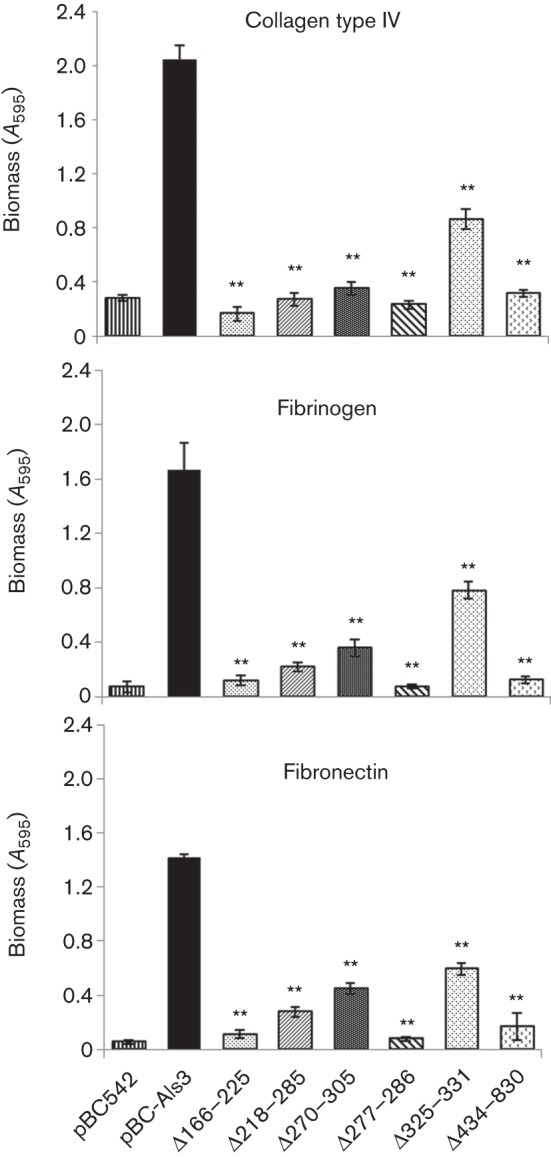
The opportunistic pathogen Candida albicans colonizes the oral cavity and gastrointestinal tract. Adherence to host cells, extracellular matrix and salivary glycoproteins that coat oral surfaces, including prostheses, is an important prerequisite for colonization. In addition, interactions of C. albicans with commensal oral streptococci are suggested to promote retention and persistence of fungal cells in mixed-species communities. The hyphal filament specific cell wall protein Als3, a member of the Als protein family, is a major determinant in C. albicans adherence. Here, we utilized site-specific in-frame deletions within Als3 expressed on the surface of heterologous Saccharomyces cerevisiae to determine regions involved in interactions of Als3 with Streptococcus gordonii. N-terminal region amino acid residue deletions Δ166-225, Δ218-285, Δ270-305 and Δ277-286 were each effective in inhibiting binding of Strep. gordonii to Als3. In addition, these deletions differentially affected biofilm formation, hydrophobicity, and adherence to silicone and human tissue proteins. Deletion of the central repeat domain (Δ434-830) did not significantly affect interaction of Als3 with Strep. gordonii SspB protein, but affected other adherence properties and biofilm formation. Deletion of the amyloid-forming region (Δ325-331) did not affect interaction of Als3 with Strep. gordonii SspB adhesin, suggesting this interaction was amyloid-independent. These findings highlighted the essential function of the N-terminal domain of Als3 in mediating the interaction of C. albicans with S. gordonii, and suggested that amyloid formation is not essential for the inter-kingdom interaction.


Figures

Fig. 1.
Fig. 1.
Diagrammatic representation of the Als3 polypeptide showing (a) overall structure and domains based upon primary sequence, (b) corresponding protein expressed on the surface of Sacch. cerevisiae with insertion of a HA-tag and replacement of the C-terminal region with an Epa1 Ser/Thr-rich stalk from Candida glabrata and (c) proteins expressed by als3 plasmid deletion mutants and expressed on the surface of Sacch. cerevisiae. Numbers indicate amino acid residues. Amino acid residues 325–331 corresponded to the predicted β-amyloid sequence IVIVATT (AFR). GPI, glycosylphosphatidylinositol.
Fig. 2.
Fig. 2.
Interactions of Sacch. cerevisiae expressing Als3 or Als3Δ polypeptides with Strep. gordonii DL1. Sacch. cerevisiae cells were incubated for 3 h at 30 °C and then with FITC-labelled Strep. gordonii for 1 h. Interactions were visualized by transmitted light microscopy and by fluorescence microscopy. (a–h) Light and corresponding fluorescence images of Sacch. cerevisiae BY4742 strains carrying pBC542 [vector-only control (Ctrl)] (a), pBC542-Als3 (b), pBC542-Als3Δ166–225 (c), pBC542-Als3Δ218–285 (d), pBC542-Als3Δ270–305 (e), pBC542-Als3Δ277–286 (f), pBC542-Als3Δ325–331 (g) and pBC542-Als3Δ434–830 (h). Bar, 50 µm.
Fig. 3.
Fig. 3.
Binding levels of Strep. gordonii DL1 to Sacch. cerevisiae expressing Als3 or Als3Δ polypeptides. Sacch. cerevisiae cells were incubated for 3 h at 30 °C and then with FITC-labelled Strep. gordonii for 1 h. Strep. gordonii adherence is expressed as mean±sd (n = 3) RFU per Sacch. cerevisiae cell, as described in Methods. Statistical significance relative to pBC-Als3: *P<0.05, **P<0.005.
Fig. 4.
Fig. 4.
Interactions of Sacch. cerevisiae expressing Als3 or Als3Δ polypeptides with surrogate host L. lactis cells expressing SspB polypeptide adhesin from Strep. gordonii. Sacch. cerevisiae cells were incubated for 3 h at 30 °C and then with FITC-labelled L. lactis for 1 h. Interactions were visualized by transmitted light microscopy and by fluorescence microscopy. (a–h) Light and corresponding fluorescence images of Sacch. cerevisiae BY4742 strains carrying pBC542 [vector-only control (Ctrl)] (a), pBC542-Als3 (b), pBC542-Als3Δ166–225 (c), pBC542-Als3Δ218–285 (d), pBC542-Als3Δ270–305 (e), pBC542-Als3Δ277–286 (f), pBC542-Als3Δ325–331 (g) and pBC542-Als3Δ434–830 (h). Bar, 50 µm.
Fig. 5.
Fig. 5.
Binding levels of L. lactis expressing Strep. gordonii SspB protein to Sacch. cerevisiae expressing Als3 or Als3Δ polypeptides. Sacch. cerevisiae cells were incubated for 3 h at 30 °C and then with FITC-labelled L. lactis for 1 h. L. lactis adherence is expressed as mean±sd (n = 3) RFU per Sacch. cerevisiae cell, as described in Methods. Statistical significance relative to pBC-Als3: **P<0.005.
Fig. 6.
Fig. 6.
Biofilm formation by Sacch. cerevisiae cells expressing Als3 or Als3Δ polypeptides. Sacch. cerevisiae cells were incubated with saliva-coated coverslips for 24 h at 30 °C. After washing and air-drying, cells were stained with crystal violet and imaged by light microscopy. (a) Crystal violet stained biofilms formed by Sacch. cerevisiae BY4742 strains carrying pBC542 [vector-only control (Ctrl)] (A), pBC542-Als3 (B), pBC542-Als3Δ166–225 (C), pBC542-Als3Δ218–285 (D), pBC542-Als3Δ270–305 (E), pBC542-Als3Δ277–286 (F), pBC542-Als3Δ325–331 (G) and pBC542-Als3Δ434–830 (H). Bar, 50 µm. (b) Corresponding biomass values for biofilms measured by crystal violet assay (see Methods). Values shown represent mean±sd of three experiments performed in triplicate. Significant differences in biomass from the Sacch. cerevisiae Als3-expressing strain: * P<0.05, **P<0.005.
Fig. 7.
Fig. 7.
Early stage biofilm formation on (a) polystyrene or (b) serum-coated silicone elastomer by Sacch. cerevisiae cells expressing Als3 or Als3Δ polypeptides, or carrying empty vector (pBC542). (a) Sacch. cerevisiae cells were incubated in polystyrene microtitre plate wells for 4 h at 30 °C. Non-adherent cells were removed and adhered biomass measured by crystal violet staining. (b) Sacch. cerevisiae cells were incubated with serum-coated silicone elastomer squares (1 cm2) for 4 h at 30 °C. Non-adherent cells were removed and total biomass was measured by crystal violet assay. Values shown represent mean±sd of three experiments performed in triplicate. Significant differences in biomass from the Sacch. cerevisiae pBC-Als3+-expressing strain: *P<0.05, **P<0.005.
Fig. 8.
Fig. 8.
Early-stage biofilm formation on ECM proteins (collagen type IV, fibrinogen or plasma fibronectin) by Sacch. cerevisiae cells expressing Als3 polypeptides. Sacch. cerevisiae cells were incubated in microwells coated with 0.5 µg ECM protein for 4 h at 30 °C. Non-adherent cells were removed and total biomass values were estimated by crystal violet staining. Values shown represent mean±sd of three experiments performed in triplicate. Significant differences in biomass from the Sacch. cerevisiae pBC-Als3+-expressing strain: **P<0.005.

References

    1. Alsteens D., Garcia M. C., Lipke P. N., Dufrêne Y. F. (2010). Force-induced formation and propagation of adhesion nanodomains in living fungal cells. Proc Natl Acad Sci U S A 107, 20744–20749. 10.1073/pnas.1013893107 - DOI - PMC - PubMed
    1. Bamford C. V., d’Mello A., Nobbs A. H., Dutton L. C., Vickerman M. M., Jenkinson H. F. (2009). Streptococcus gordonii modulates Candida albicans biofilm formation through intergeneric communication. Infect Immun 77, 3696–3704. 10.1128/IAI.00438-09 - DOI - PMC - PubMed
    1. Beaussart A., Alsteens D., El-Kirat-Chatel S., Lipke P. N., Kucharíková S., Van Dijck P., Dufrêne Y. F. (2012). Single-molecule imaging and functional analysis of Als adhesins and mannans during Candida albicans morphogenesis. ACS Nano 6, 10950–10964. - PMC - PubMed
    1. Brachmann C. B., Davies A., Cost G. J., Caputo E., Li J., Hieter P., Boeke J. D. (1998). Designer deletion strains derived from Saccharomyces cerevisiae S288C: a useful set of strains and plasmids for PCR-mediated gene disruption and other applications. Yeast 14, 115–132. 10.1002/(SICI)1097-0061(19980130)14:2<115::AID-YEA204>3.0.CO;2-2 - DOI - PubMed
    1. Cannon R. D., Chaffin W. L. (1999). Oral colonization by Candida albicans. Crit Rev Oral Biol Med 10, 359–383. 10.1177/10454411990100030701 - DOI - PubMed

Publication types

Substances

LinkOut - more resources