. 2014 Nov 10;42(20):12498-511.
doi: 10.1093/nar/gku995. Epub 2014 Oct 20.

N-terminal phosphorylation of HP1α increases its nucleosome-binding specificity

Affiliations

N-terminal phosphorylation of HP1α increases its nucleosome-binding specificity

Gohei Nishibuchi et al. Nucleic Acids Res. .

Abstract

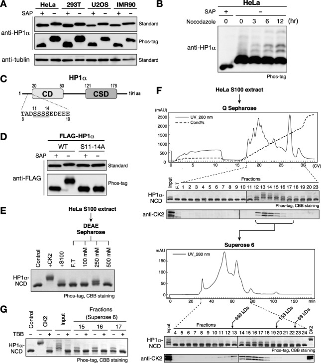
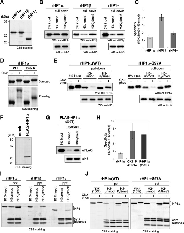
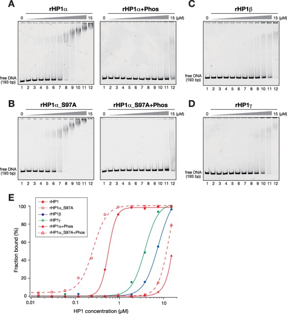
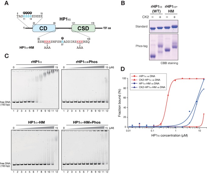
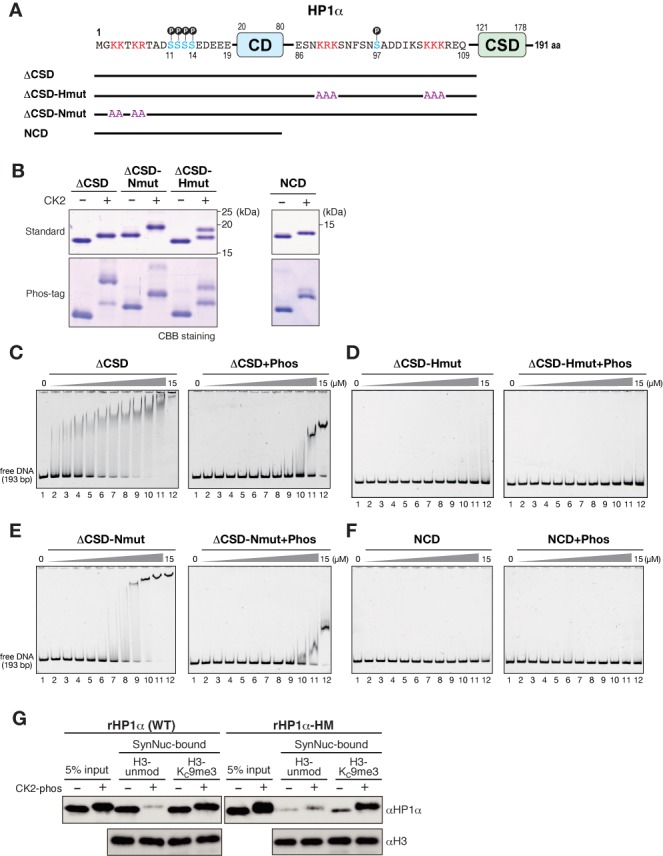
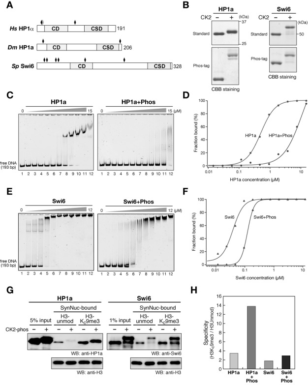
Heterochromatin protein 1 (HP1) is an evolutionarily conserved chromosomal protein that binds to lysine 9-methylated histone H3 (H3K9me), a hallmark of heterochromatin. Although HP1 phosphorylation has been described in several organisms, the biological implications of this modification remain largely elusive. Here we show that HP1's phosphorylation has a critical effect on its nucleosome binding properties. By in vitro phosphorylation assays and conventional chromatography, we demonstrated that casein kinase II (CK2) is the kinase primarily responsible for phosphorylating the N-terminus of human HP1α. Pull-down assays using in vitro-reconstituted nucleosomes showed that unmodified HP1α bound H3K9-methylated and H3K9-unmethylated nucleosomes with comparable affinity, whereas CK2-phosphorylated HP1α showed a high specificity for H3K9me3-modified nucleosomes. Electrophoretic mobility shift assays showed that CK2-mediated phosphorylation diminished HP1α's intrinsic DNA binding, which contributed to its H3K9me-independent nucleosome binding. CK2-mediated phosphorylation had a similar effect on the nucleosome-binding specificity of fly HP1a and S. pombe Swi6. These results suggested that HP1 phosphorylation has an evolutionarily conserved role in HP1's recognition of H3K9me-marked nucleosomes.


Figures

Figure 1.
Figure 1.
HP1α is constitutively phosphorylated by CK2 at N-terminal serine residues in human cells. (A) Phosphorylation states of HP1α in various human cell lines. SAP-treated samples were used as unphosphorylated basal control proteins. Samples were resolved on 12% standard SDS-PAGE gel (standard) or on 10% SDS-PAGE gel containing 50 μM Phos-tag (Phos-tag) gel, and analyzed by western blotting with an anti-HP1α antibody. Western blotting using an anti-α-tubulin antibody is shown for a loading control. (B) Phosphorylation patterns of human HP1α in G2/M-arrested cells. HeLa cells treated with 200 ng/ml nocodazole for 3, 6 or 12 h were harvested and analyzed as in (A). (C) Schematic diagram of the HP1α protein and its N-terminal amino acid sequence. Serine residues that are phosphorylatable by CK2 are underlined. (D) Serine clusters (S11–14) were constitutively phosphorylated in vivo. FLAG-tagged WT HP1α or HP1α with a S11–14A mutation was transiently expressed in HEK293T cells and analyzed as in (A). (E) In vitro kinase assays using recombinant protein containing the HP1α N-terminus and CD (HP1α-NCD). HeLa cytoplasmic extract (S100) and fractions that were partially purified through DEAE Sepharose were assayed. (F) Partial purification of cellular kinase(s) that phosphorylate HP1α-NCD: HeLa S100 extracts were loaded onto a HiTrap Q HP column, and each fraction was subjected to a kinase assay (top panels). Fractions containing kinase activity were pooled and further separated on a Superose 6 Column (bottom panels). Elution profiles of endogenous CK2 were analyzed by western blotting using an anti-CK2α antibody (beneath each panel). (G) Representative fractions in (E) were subjected to the in vitro kinase assay in the presence or absence of the CK2-specific inhibitor TBB.
Figure 2.
Figure 2.
The N-terminal phosphorylation of HP1α increases its binding specificity for H3K9me3 nucleosomes. (A) Recombinant human HP1α, HP1β and HP1γ prepared from E. coli. (B) Representative nucleosome pull-down assays using synthesized nucleosomes containing unmodified H3 (H3unmod) or H3 with a K9me3 analog (H3Kc9me3), and biotinylated 193-bp 601 DNA. Recombinant human HP1α, HP1β or HP1γ was incubated with nucleosomes immobilized on streptavidin-beads, and the input and recovered proteins were resolved by SDS-PAGE and immunoblotted using cognate antibodies. (C) Quantified ratios of H3Kc9me3 nucleosome-bound HP1 over HP1 bound to H3unmod nucleosomes. Error bars represent the range of the standard deviations of three independent measurements. (D) Control and CK2-phosphorylated WT and S97A HP1α proteins were resolved by standard or Phos-tag PAGE. (E) Representative nucleosome pull-down assays using control and CK2-phosphorylated HP1αs. (F) FLAG-tagged HP1α (FLAG-HP1α) prepared from HEK293T. FLAG-HP1α transiently expressed in HEK293T cells was purified using anti-FLAG M2 agarose. A mock purification result is also shown (mock). (G) A representative nucleosome pull-down assay using HP1α prepared from HEK293T cells. (H) The quantified ratios of H3Kc9me3 nucleosome-bound HP1α over HP1α bound to H3unmod nucleosomes. Error bars represent the range of the standard deviations of three independent measurements. (I and J) Chromatin co-precipitation assays using 12-mer nucleosomal arrays. Recovered nucleosomes and bound HP1 proteins were analyzed by15% SDS-PAGE and visualized by CBB staining.
Figure 3.
Figure 3.
HP1α's N-terminal phosphorylation inhibits its DNA binding. (A and B) Representative EMSAs using control and CK2-phosphorylated, recombinant HP1α (rHP1α and rHP1α+Phos) (A) and HP1α with S97A mutations (rHP1α_S97A and rHP1α_S97A+Phos) (B). The HP1 concentration varied from 0 to 15 μM (0.5-fold dilutions). A 193-bp 601 DNA was used as a probe. Retarded probes in native PAGE gels were stained with SYBR Gold and detected by LAS3000. (C and D) Recombinant HP1β (C) and HP1γ (D) were subjected to EMSA as described in (A). (E) Quantification of EMSAs using HP1 proteins. The fraction of retarded DNA probe, determined by EMSA (A–D), was plotted against the HP1 protein concentration.
Figure 4.
Figure 4.
HP1α's N-terminal phosphorylation inhibits its hinge region-mediated DNA binding. (A) Schematic diagram of HP1α showing amino acid sequences of the N-terminal and hinge regions, and the positions of amino acid substitutions in an HP1α with a hinge mutation (HP1α-HM). Phosphorylatable serine residues are shown in blue, and the basic residues replaced by alanine in the HP1α-HM are shown in red. (B) Control and CK2-phosphorylated WT and hinge-mutated (HM) HP1α proteins were resolved by standard or Phos-tag PAGE and visualized by CBB staining. (C) Representative EMSAs using control and CK2-phosphorylated WT and HM HP1α. The HP1 concentration varied from 0 to 15 μM (0.5-fold dilutions). A 193-bp 601 DNA was used as a probe. (D) Quantification of EMSAs using HP1 proteins. The fraction of retarded DNA probe, determined by EMSA (C), was plotted against the HP1 protein concentration.
Figure 5.
Figure 5.
Basic amino acid residues at HP1α's N-terminal tail modulate DNA binding involving the hinge region. (A) Schematic diagram of full-length HP1α with the amino acid sequences of the N-terminal and hinge regions (top), and truncated ΔCSD mutants with amino acid substitutions. Phosphorylatable serine residues are shown in blue, and the basic residues replaced by alanine in hinge-mutated (Hmut) or N-terminal-mutated (Nmut) ΔCSD constructs are shown in red. (B) Control and CK2-phosphorylated, ΔCSD, ΔCSD-Nmut, CSD-Hmut and NCD proteins were resolved by standard or Phos-tag PAGE and visualized by CBB staining. (C–F) Representative EMSAs using control and CK2-phosphorylated ΔCSD (C), ΔCSD-Hmut (D), ΔCSD-Nmut and NCD mutants (F). The protein concentration varied from 0 to 15 μM (0.5-fold dilutions). A 193-bp 601 DNA was used as a probe. (G) Nucleosome pull-down assays using synthesized nucleosomes. Recombinant WT HP1α or HP1α with a HM were incubated with nucleosomes immobilized on streptavidin-beads, and the input and recovered proteins were resolved by SDS-PAGE and immunoblotted using an anti-HP1α antibody.
Figure 6.
Figure 6.
Conserved role of CK2-mediated phosphorylation in HP1's nucleosome binding. (A) Schematic drawings of human (Hs) HP1α, Drosophila (Dm) HP1a and S. pombe (Sp) Swi6. The positions of CK2-phosphorylatable serine residues are indicated by black rhomboids. (B) Control and CK2-phosphorylated HP1a and Swi6 were resolved by standard or Phos-tag PAGE and visualized by CBB staining. (C and E) Representative EMSAs using control and CK2-phosphorylated HP1a (C) and Swi6 (E). The protein concentration varied from 0 to 15 μM for HP1a, and from 0 to 12 μM for Swi6, respectively (0.5-fold dilutions). A 193-bp 601 DNA was used as a probe. (D and F) Quantification of EMSAs using HP1 proteins. The fraction of retarded DNA probe in (C) and (E) was plotted against the HP1 protein concentration. (G) Nucleosome pull-down assays using synthesized nucleosomes. Control and CK2-phosphorylated HP1a (left) and Swi6 (right) were incubated with nucleosomes immobilized on streptavidin beads, and the input and recovered proteins were resolved by SDS-PAGE and immunoblotted using cognate antibodies. (H) The quantified ratios of H3Kc9me3 nucleosome-bound HP1a or Swi6 over that bound to unmodified nucleosomes.

References

    1. Grewal S.I., Elgin S.C. Heterochromatin: new possibilities for the inheritance of structure. Curr. Opin. Genet. Dev. 2002;12:178–187. - PubMed
    1. Richards E.J., Elgin S.C. Epigenetic codes for heterochromatin formation and silencing: rounding up the usual suspects. Cell. 2002;108:489–500. - PubMed
    1. Grewal S.I., Moazed D. Heterochromatin and epigenetic control of gene expression. Science. 2003;301:798–802. - PubMed
    1. Bannister A.J., Zegerman P., Partridge J.F., Miska E.A., Thomas J.O., Allshire R.C., Kouzarides T. Selective recognition of methylated lysine 9 on histone H3 by the HP1 chromo domain. Nature. 2001;410:120–124. - PubMed
    1. Lachner M., O'Carroll D., Rea S., Mechtler K., Jenuwein T. Methylation of histone H3 lysine 9 creates a binding site for HP1 proteins. Nature. 2001;410:116–120. - PubMed

Publication types