Review
. 2014 Nov 10;42(20):12352-66.
doi: 10.1093/nar/gku999. Epub 2014 Oct 20.

G-quadruplexes in viruses: function and potential therapeutic applications

Affiliations
Review

G-quadruplexes in viruses: function and potential therapeutic applications

Mathieu Métifiot et al. Nucleic Acids Res. .

Abstract

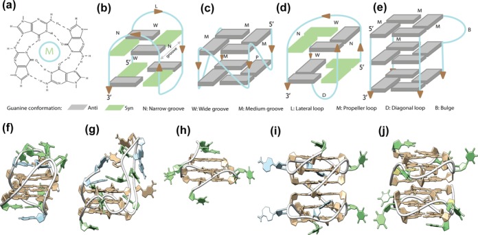
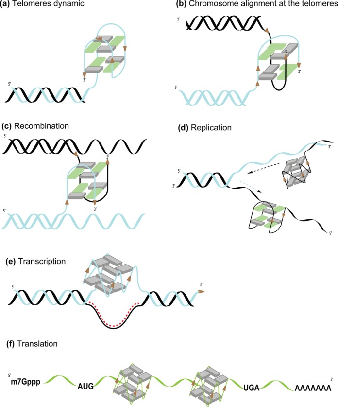
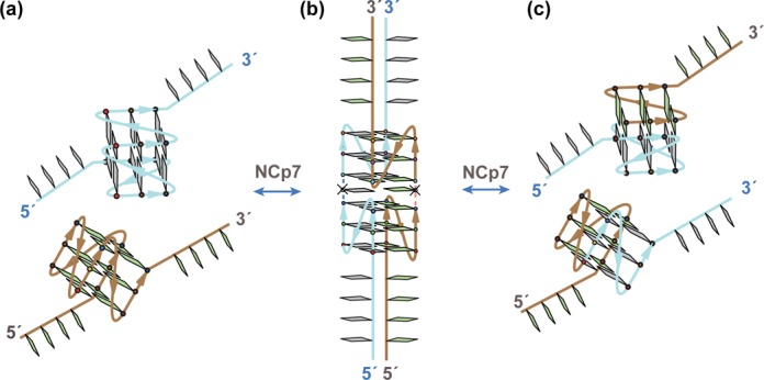
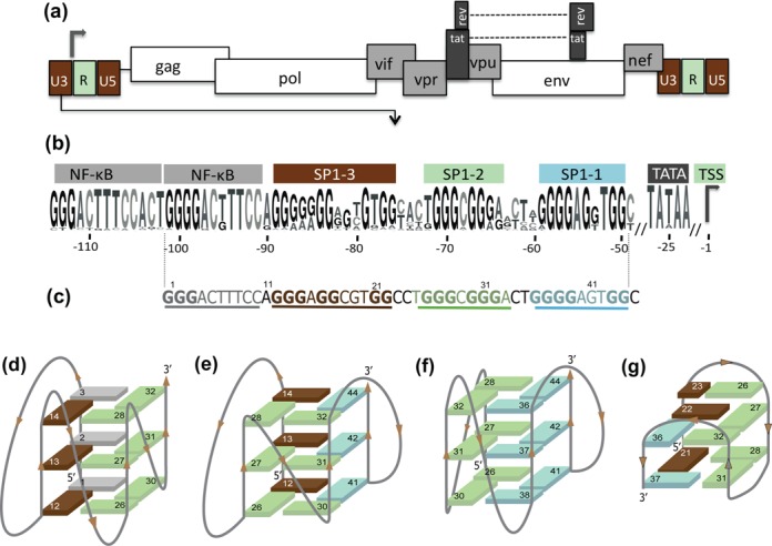
G-rich nucleic acids can form non-canonical G-quadruplex structures (G4s) in which four guanines fold in a planar arrangement through Hoogsteen hydrogen bonds. Although many biochemical and structural studies have focused on DNA sequences containing successive, adjacent guanines that spontaneously fold into G4s, evidence for their in vivo relevance has recently begun to accumulate. Complete sequencing of the human genome highlighted the presence of ∼300,000 sequences that can potentially form G4s. Likewise, the presence of putative G4-sequences has been reported in various viruses genomes [e.g., Human immunodeficiency virus (HIV-1), Epstein-Barr virus (EBV), papillomavirus (HPV)]. Many studies have focused on telomeric G4s and how their dynamics are regulated to enable telomere synthesis. Moreover, a role for G4s has been proposed in cellular and viral replication, recombination and gene expression control. In parallel, DNA aptamers that form G4s have been described as inhibitors and diagnostic tools to detect viruses [e.g., hepatitis A virus (HAV), EBV, cauliflower mosaic virus (CaMV), severe acute respiratory syndrome virus (SARS), simian virus 40 (SV40)]. Here, special emphasis will be given to the possible role of these structures in a virus life cycle as well as the use of G4-forming oligonucleotides as potential antiviral agents and innovative tools.


Figures

Figure 1.
Figure 1.
(ae) Schematic representation of G4 topologies. (a) A guanine tetrad stabilized by eight Hoogsteen hydrogen bonds and a central monovalent cation (M). (b) Intramolecular antiparallel G4 topology with two tetrads, wide and narrow grooves and only lateral loops. (c) Intramolecular parallel G4 topology with two tetrads, medium grooves and only propeller loops. (d) Dimeric antiparallel G4 topology with two tetrads, wide and narrow grooves and diagonal loops. (e) Tetramolecular parallel G4 topology with three tetrads, only medium grooves and no loops. (fj) Examples of G4 structures. (f) Intramolecular anti-parallel G4 structure with two tetrads for the telomeric sequence (PDB ID: 2KF8). (g) Intramolecular parallel G4 structure with three tetrads and a nine nucleotide central loop for the human CEB25 mini-satellite sequence (PDB ID: 2LPW). (h) Intramolecular parallel G4 structure with three tetrads for the T30177 anti-HIV aptamer (PDB ID: 2M4P). (i) Interlocked bimolecular parallel G4 structure with six tetrads for the 93del anti-HIV aptamer (PDB ID: 1Y8D). (j) Two stacked parallel G4 structures with three tetrads each observed for the T30923 anti-HIV aptamer (PDB ID: 2LE6).
Figure 2.
Figure 2.
Role of G4 structures in the genome. (a) Formation of a G4 at the 3′ end of a chromosome. (b) Formation of an intermolecular G4 between two telomeres allows chromosomes’ alignment. (c) Intermolecular G4 formation leads to physical crossing and potential recombination sites at different locations of the DNA (d) Collision of the replication fork with G4s formed on the leading and lagging DNA strands. (e) Transcription can lead to the formation of a DNA/RNA duplex (R-loop) on the template strand and formation of a G4 (G-loop) on the non-template strand. (f) Formation of G4s within mRNAs block translation, as observed for EBNA1 coding mRNA (131).
Figure 3.
Figure 3.
Model of viral RNA:RNA dimerization and recombination involving a pair of monomeric G4s mediated by a synaptic dimeric G4 intermediate [inspired and adapted from a model of intergenic recombination (115)]. (a) A pair of parallel-stranded monomeric G4s. (b) NCp7 chaperon properties favor formation of an inter-RNA synaptic intermediate involving a parallel-stranded dimeric G4. Cleavage, rotation and subsequent rejoining are mediated by a potential, undefined nuclease-topoisomerase-like enzyme complex. (c) Following dimerization and/or recombination, NCp7 annealing properties favor formation of a new pair of parallel-stranded monomeric G4s.
Figure 4.
Figure 4.
(a) Genomic structure of the HIV-1 provirus and (b) LOGO representation of the G-rich region for the HIV-1 promoter generated using the weblogo software (183) and based on an alignment of 1684 HIV-1 sequences provided by the HIV-1 database (www.hiv.lanl.gov). (c) The U3 region of the LTR from the HXB2_LAI;NC_001802 HIV-1 representative strain. (d, e and g) Topologies of the LTR G4s determined using Clerocidin and DMS-mediated footprinting assays (116) for the (d) 1–32 nt segment, (e) 12–44 nt segment and (f) 26–44 nt segment. (g) Topology of the 12–37 nt segments determined using nuclear magnetic resonance (NMR) (117).

References

    1. Bang I. Untersuchungen über die Guanylsäure. Biochem. Z. 1910;26:293–311.
    1. Gellert M., Lipsett M.N., Davies D.R. Helix formation by guanylic acid. Proc. Natl. Acad. Sci. U.S.A. 1962;48:2013–2018. - PMC - PubMed
    1. Sasisekharan V., Zimmerman S., Davies D.R. The structure of helical 5′-guanosine monophosphate. J. Mol. Biol. 1975;92:171–179. - PubMed
    1. Howard F.B., Miles H.T. Poly(inosinic acid) helices: essential chelation of alkali metal ions in the axial channel. Biochemistry. 1982;21:6736–6745. - PubMed
    1. Williamson J.R. Guanine quartets. Curr. Opin. Struct. Biol. 1993;3:357–362.

Publication types