Eosinophilic vasculitis: time for recognition of a new entity?
- PMID: 25332610
- PMCID: PMC4192264
- DOI: 10.1007/s12288-014-0384-2
Eosinophilic vasculitis: time for recognition of a new entity?
Abstract
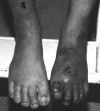
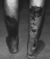
Hypereosinophilia is part of a group of complex disorders with multisystem involvement. A 23 year old male was admitted to our centre with bilateral popliteal artery and venous thrombosis and impending gangrene of the left forefoot along with deep venous thrombosis of the right lower extremity. Investigations revealed marked peripheral blood eosinophilia (27,669/μL). Bone marrow showed increased eosinophils & eosinophil precursors and no evidence of a clonal disorder. Skin biopsy from the ulcerated lesions showed small vessel vasculitis with intense eosinophilic infiltration. Investigations to look for secondary causes of hypereosinophilia in the form of Antinuclear Antibody, P-Anti Neutrophil Cytoplasmic Antibody (ANCA) and C-ANCA and FIP1L1-PDGFRA, Bcr-Abl and JAK2V617F mutations were negative. The arterial and venous thrombosis and cutaneous vasculitis were linked to the presence of hypereosinophilic syndrome. The patient's illness responded to high dose corticosteroids leading to complete resolution of symptoms. We reviewed the literature on the lesser known entity of eosinophilic vasculitis and its association with thrombosis.
Keywords: Eosinophilia; Thrombosis; Vasculitis.
Figures


References
-
- Liao YH, Su YW, Tsay W, Chiu HC. Association of cutaneous necrotizing eosinophilic vasculitis and deep vein thrombosis in hypereosinophilic syndrome. Arch Dermatol. 2005;141(8):1051–1053. - PubMed
Publication types
LinkOut - more resources
Full Text Sources
Other Literature Sources
Miscellaneous
