. 2014 Jun 6;1(1):e000020.
doi: 10.1136/openhrt-2013-000020. eCollection 2014.

Percutaneous left atrial appendage occlusion for stroke prevention in atrial fibrillation: an update

Affiliations

Percutaneous left atrial appendage occlusion for stroke prevention in atrial fibrillation: an update

O De Backer et al. Open Heart. .

Abstract

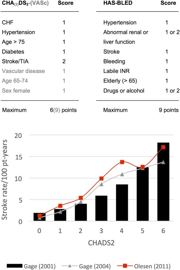
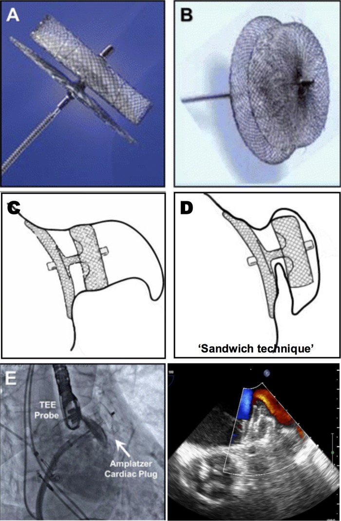
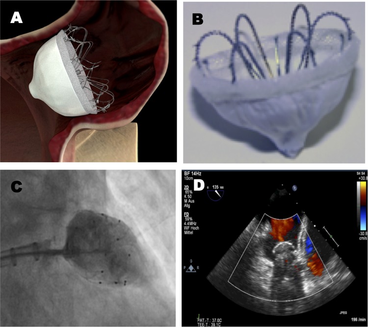
Atrial fibrillation (AF) is the most common sustained cardiac arrhythmia encountered in clinical practice. One of its most devastating complications is the development of thromboembolism leading to fatal or disabling stroke. Oral anticoagulation (OAC, warfarin) is the standard treatment for stroke prevention in patients with AF with an increased stroke risk. However, there are several obstacles to long-term OAC therapy, including the risk of serious bleeding, several drug-drug interactions and the need for frequent blood testing. Although newer oral anticoagulants have been developed, these drugs also face issues of major bleeding and non-compliance. Therefore, alternative treatment options for stroke prevention in patients with AF with a high stroke risk are needed. Percutaneous left atrial appendage (LAA) occlusion is an evolving therapy, which should be taken into consideration in those patients with non-valvular AF with a high stroke risk and contraindications for OAC. This article aims to discuss the rationale for LAA closure, the available LAA occlusion devices and their clinical evidence until now. Moreover, we discuss the importance of proper patient selection, the role of various imaging techniques and the need for a more tailored postprocedural antithrombotic therapy.

Keywords: Allied Specialities.


Figures

Figure 1
Figure 1
The CHA(2)DS2-(VASc) stroke risk and HAS-BLED bleeding risk index are calculated by totalling the scores for each risk factor present. The lower graph shows the expected stroke rate /100 patient (pt)-years, stratified by CHADS2 score in patients with AF not taking warfarin. Gage (2001): adjusted stroke rates/100 pt-years, assuming that aspirin was not taken; Gage (2004): stroke rates/100 pt-years of aspirin; Olesen (2011): event rates of hospital admission and death due to thromboembolism in patients with AF not taking warfarin. AF, atrial fibrillation; CHF, congestive heart failure; TIA, transient ischaemic attack; INR, international normalised ratio.
Figure 2
Figure 2
General morphology classification of left atrial appendage (LAA) as determined by cardiac CT. LAA can be classified into four types: (A) chicken wing type—with a short neck and an obvious bend, (B) windsock type, (C) cauliflower type and (D) cactus type. Images reproduced with permission from Wang et al.
Figure 3
Figure 3
(A–D) Preprocedural transoesophageal echocardiography (TEE) imaging—left atrial appendage (LAA) anatomy and dimensions should be studied with the transducer array rotated through 0°, 45°, 90° and 135°. (A) Shows in which manner the LAA ostium, LAA neck width (orifice width or ‘landing zone’) and LAA depth should be measured. The LAA neck width is typically measured in a plane from the LCx coronary artery to a point 10 mm distal to the limbus (white arrow) of the left superior pulmonary vein (LSPV). In this case, we measured a neck width of 21–25 mm—which led to the choice of a 28 mm Amplatzer cardiac plug (ACP) device (because of oversizing with 3 to 5 mm). The risk of undersizing is device embolisation; the risk of oversizing is compression on the LCx and/or LSPV, as well as LAA perforation and device embolisation. (E–H) Use of ICE to guide implantation of a LAA closure device. ICE imaging is optimal with the ICE probe in (E) the left pulmonary artery as well as (G) with the transducer at the ostium of the coronary sinus. The ICE images were used to guide (F) the delivery and (H) proper deployment of the ACP device inside the LAA. DC, delivery cable; *disk of the ACP device; # lobe of the ACP device.
Figure 4
Figure 4
PLAATO device. (A) The PLAATO (Percutaneous Left Atrial Appendage Transcatheter Occlusion) system was the first device specifically developed for left atrial appendage (LAA) occlusion. It consisted of a self-expanding nitinol cage with three anchors on each strut and was covered with a non-thrombogenic PTFE membrane. The anchoring barbs provided the stability; the PTFE membrane prevented mobilisation of thrombi from the LAA and promoted healing. The device diameter ranged between 15 and 32 mm and was normally selected 20–40% larger than the diameter of the LAA ostium. The device is no longer available for clinical use after withdrawal from the market in 2006. (B) Shows—from left to right—a fully collapsed, partially expanded and fully expanded device advanced through a 12-Fr transseptal delivery sheath. (C, D) Illustrate a fluoroscopic right anterior oblique view in a patient, exhibiting the deployed PLAATO device (C) and in the setting of an LA angiogram (D) depicting proper LAA occlusion. Images reproduced with permission from Aryana et al.
Figure 5
Figure 5
WATCHMAN device. (A–C) Show the delivery (A), deployment (B), and release (C) of the WATCHMAN device through a 12-Fr transseptal delivery sheath. (D) Shows a close-up view of the WATCHMAN device—consisting of a self-expanding nitinol frame covered with a porous filtering PET membrane. The stability of the device is secured by fixation barbs located circumferentially; the PET membrane acts as a filter preventing the outflow of the thrombi and promotes endothelialisation. The device is available in five different sizes ranging from 21 to 33 mm, and is normally selected 10–20% larger than the left atrial appendage (LAA) diameter to ensure stable device positioning. The device can be recaptured and withdrawn in case of suboptimal fixation. The WATCMAN device received CE-mark approval in 2005 and is currently used in clinical practice. (E) Shows a transoesophageal echocardiography image of an occluded LAA following deployment of a WATCHMAN device—the delivery cable is still connected to the device. (F) Shows a cine image of an LA angiogram demonstrating a WATCHMAN device properly deployed inside the LAA (black arrow). TSS, transseptal sheath. Images reproduced with permission from Aryana et al.
Figure 6
Figure 6
(A-B) the ACP device consists of a lobe and a disk connected by a short, flexible waist. Both the lobe and disk are constructed from a nitinol mesh covered with a polyester patch. The lobe is implanted within the neck of the LAA (the so-called ‘landing zone’), and achieves device stabilization and retention by means of a number of stabilization wires. The delivery system is 9-13Fr depending on the size of the device. The lobe size ranges from 16-30 mm and the disk from 20–36 mm; the size of the lobe should be chosen 3 to 5 mm larger than the diameter of the ‘landing zone’. The ACP device is not designed to fill the LAA but to seal its ostium by means of the larger disk. As such, the ACP device could be a better choice when challenged with a more complex anatomy of the distal LAA or a proximal LAA lobe. The ACP device received a CE mark in 2008 and is currently used in clinical practice. (C-D) show the implantation of an ACP device in the regular way (C), or using the ‘sandwich technique’ when confronted with a chicken wing LAA with a short neck (D). (E) a cine image of a LA angiogram performed through a transseptal catheter following deployment of an ACP device (white arrow) inside the LAA - when properly positioned, the lobe has a typical ‘tire’ morphology with slight compression on the sides. (F) a TEE image of a properly deployed ACP device, showing absence of peri-device leaks, good alignment of the disk with the LA cavity, and absence of compression on the left upper pulmonary vein. Some images were reproduced with permission from Aryana et al. (2012).
Figure 7
Figure 7
(A-B) the Coherex WaveCrest LAA occlusion system is the latest development in LAA occlusion devices. It consists of a nitinol frame with retractable coils and anchors to enable optimal device positioning. The device consists of a multi-composite membrane including a PTFE membrane on the LA side of the device, and a foam substrate on the LA-opposing surface to minimize residual leaks. Very recently, the WAVECREST I clinical trial was completed - CE Mark approval was obtained in September 2013. (C) a cine image showing contrast injection distally of the device (into the LAA) in order to assess complete LAA closure. (D) a TTE image confirming the device is well-seated and without leaks. Some images were presented by Franzen O. on EuroPCR 2013.

References

    1. Wolf PA, Abbott RD, Kannel WB. Atrial fibrillation as an independent risk factor for stroke: the Framingham Study. Stroke 1991;22:983–8 - PubMed
    1. Furberg CD, Psaty BM, Manolio TA, et al. Prevalence of atrial fibrillation in elderly subjects—the Cardiovascular Health Study. Am J Cardiol 1994;74:236–41 - PubMed
    1. Go AS, Hylek EM, Philips KA, et al. Prevalence of diagnosed atrial fibrillation in adults: national implications for rhythm management and stroke prevention: the AnTicoagulation and Risk Factors in Atrial Fibrillation (ATRIA) Study. JAMA 2001;285:2370–5 - PubMed
    1. Feinberg WM, Blackshear JL, Laupacis , et al. Prevalence, age distribution, and gender of patients with atrial fibrillation. Analysis and implications. Arch Intern Med 1995;155:469–73 - PubMed
    1. (No Author used). Risk factors for stroke and efficacy of antithrombotic therapy in atrial fibrillation. Analysis of pooled data from five randomized controlled trials. Arch Intern Med 1994;154:1449–57 - PubMed