. 2014 Oct 14:2:e619.
doi: 10.7717/peerj.619. eCollection 2014.

The structure of people's hair

Affiliations

The structure of people's hair

Fei-Chi Yang et al. PeerJ. .

Abstract

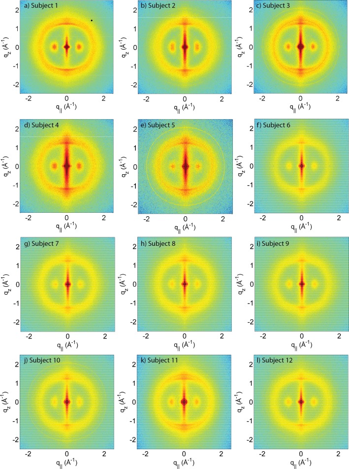
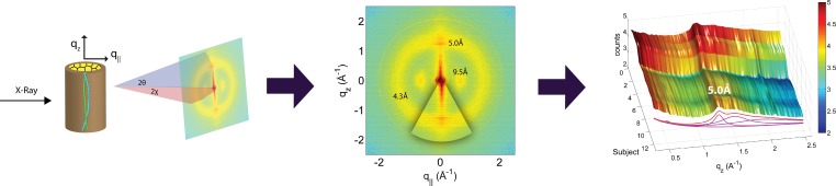
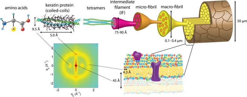
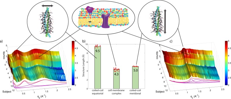
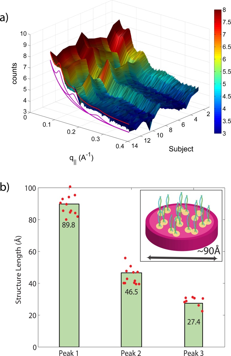
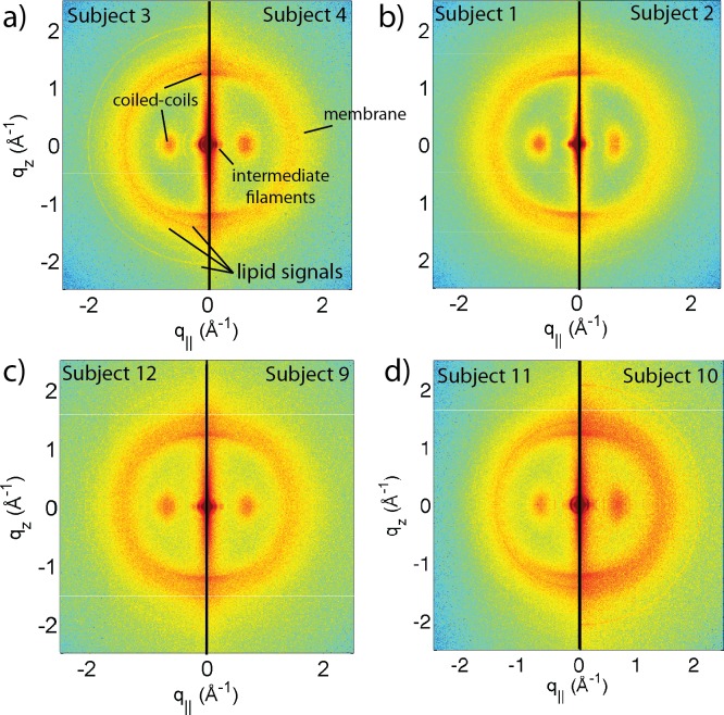
Hair is a filamentous biomaterial consisting mainly of proteins in particular keratin. The structure of human hair is well known: the medulla is a loosely packed, disordered region near the centre of the hair surrounded by the cortex, which contains the major part of the fibre mass, mainly consisting of keratin proteins and structural lipids. The cortex is surrounded by the cuticle, a layer of dead, overlapping cells forming a protective layer around the hair. The corresponding structures have been studied extensively using a variety of different techniques, such as light, electron and atomic force microscopes, and also X-ray diffraction. We were interested in the question how much the molecular hair structure differs from person to person, between male and female hair, hair of different appearances such as colour and waviness. We included hair from parent and child, identical and fraternal twins in the study to see if genetically similar hair would show similar structural features. The molecular structure of the hair samples was studied using high-resolution X-ray diffraction, which covers length scales from molecules up to the organization of secondary structures. Signals due to the coiled-coil phase of α-helical keratin proteins, intermediate keratin filaments in the cortex and from the lipid layers in the cell membrane complex were observed in the specimen of all individuals, with very small deviations. Despite the relatively small number of individuals (12) included in this study, some conclusions can be drawn. While the general features were observed in all individuals and the corresponding molecular structures were almost identical, additional signals were observed in some specimen and assigned to different types of lipids in the cell membrane complex. Genetics seem to play a role in this composition as identical patterns were observed in hair from father and daughter and identical twins, however, not for fraternal twins. Identification and characterization of these features is an important step towards the detection of abnormalities in the molecular structure of hair as a potential diagnostic tool for certain diseases.

Keywords: Alpha helix; Cell membrane complex; Coiled-coil proteins; Human hair; Intermediate filament; Keratin; Molecular structure; X-ray diffraction.


Figures

Figure 1
Figure 1. Two-dimensional X-ray data of all 12 subjects.
The hair strands were oriented with the long axis of the hair parallel to the vertical z-axis. The (q, qz)-range shown was determined in preliminary experiments to cover the features observable by X-ray diffraction. The measurements cover length scales from about 3–90 Å to study features from the coiled-coil α-keratin phase, keratin intermediate filaments in the cortex, and the membrane layer in the membrane complex. While common features can easily be identified in the 2D plots, subtle differences are visible, which are discussed in detail in the text.
Figure 2
Figure 2. The apparatus used to mount the hair strands in the experiment.
The cardboard apparatus is mounted vertically onto the loading plate of the Biological Large Angle Diffraction Experiment (BLADE) using sticky putty.
Figure 3
Figure 3. Schematics of the X-ray setup and example X-ray data.
The hair strands were oriented in the X-ray diffractometer with their long axis along qz. Two-dimensional X-ray data were measured for each specimen covering distances from about 3–90 Å including signals from the coiled-coil α-keratin phase, the intermediate fibrils in the cortex and from the cell membrane complex. The 2-dimensional data were integrated and converted into line scans and fit for a quantitative analysis.
Figure 4
Figure 4. The hierarchical structure of hair in the cortex and cuticle.
The main component of the cortex is a keratin coiled-coil protein phase. The proteins form intermediate filaments, which then organize into larger and larger fibres. The hair is surrounded by the cuticle, a dead cell layer. The common features observed in the X-ray data of all specimens are signals related to the coiled-coil keratin phase and the formation of intermediate filaments in the cortex, and the cell membrane complex. Signal assignment and corresponding length scales are shown in the figure.
Figure 5
Figure 5
Integration of the 2-dimensional scattering data in Fig. 1 in the equatorial plane (q) (A), and along the axis of the hairs (qz) (C), respectively, for all subjects. The two signals present in all individuals in the equatorial plane (q) correspond to the distance between two coiled coils of 9.5 Å and between two lipid tails in the cell membrane cortex of 4.3 Å. The common meridional signal along the long axis of the hair (qz) at 5 Å corresponds corresponds to the α-helices twisting around each other within coiled-coils. Average values and standard deviations are in (B).
Figure 6
Figure 6. Diffraction features at small scattering angles.
The small q-range is shown in magnification in (A). The specimen of most individuals showed 3 distinct reflections at ∼90 Å, 46.5 Å and 27 Å, related to the properties of intermediate keratin filaments (B).
Figure 7
Figure 7. Comparison between hair samples.
(A) shows a comparison between individuals 3 and 4. While the two specimens both show the general features, differences are observed in the region of signal from the cell membrane complex. (B) Comparison between individuals 1 and 2, father and daughter. The data in (C) (individuals 9 and 12) are from identical twins. Data in (D) was taken from fraternal twins (individuals 10 and 11). While different individuals in general show different membrane patterns (A), features in (B) and (C) perfectly agree. Fraternal twins show slight differences in their pattern in (D).

References

    1. Armstrong CL, Marquardt D, Dies H, Kučerka N, Yamani Z, Harroun TA, Katsaras J, Shi A-C, Rheinstädter MC. The observation of highly ordered domains in membranes with cholesterol. PLOS ONE. 2013;8:e619. doi: 10.1371/journal.pone.0066162. - DOI - PMC - PubMed
    1. Astbury WT, Sisson WA. X-ray studies of the structure of hair, wool, and related fibres. III. The configuration of the keratin molecule and its orientation in the biological cell. Proceedings of the Royal Society of London. Series A, Mathematical and Physical Sciences. 1935;150:533–551. doi: 10.1098/rspa.1935.0121. - DOI
    1. Astbury WT, Street A. X-ray studies of the structure of hair, wool, and related fibres. I. General. Philosophical Transactions of the Royal Society of London. Series A, Containing Papers of a Mathematical or Physical Character. 1932;230:75–101. doi: 10.1098/rsta.1932.0003. - DOI
    1. Astbury WT, Woods HJ. X-ray studies of the structure of hair, wool, and related fibres. II. The molecular structure and elastic properties of hair keratin. Philosophical Transactions of the Royal Society of London. Series A, Containing Papers of a Mathematical or Physical Character. 1934;232:333–394. doi: 10.1098/rsta.1934.0010. - DOI
    1. Barrett MA, Zheng S, Roshankar G, Alsop RJ, Belanger RKR, Huynh C, Kučerka N, Rheinstädter MC. Interaction of aspirin (acetylsalicylic acid) with lipid membranes. PLoS ONE. 2012;7:e619. doi: 10.1371/journal.pone.0034357. - DOI - PMC - PubMed