. 2014 Sep 23:3:548.
doi: 10.1186/2193-1801-3-548. eCollection 2014.

Infrared pupillometry, the Neurological Pupil index and unilateral pupillary dilation after traumatic brain injury: implications for treatment paradigms

Affiliations

Infrared pupillometry, the Neurological Pupil index and unilateral pupillary dilation after traumatic brain injury: implications for treatment paradigms

Jefferson William Chen et al. Springerplus. .

Abstract

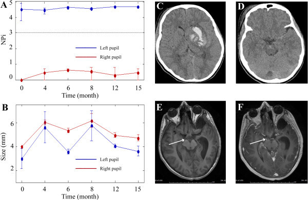
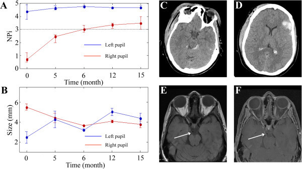
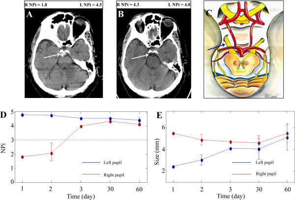
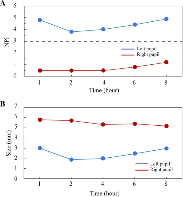
Pupillary dysfunction, a concerning finding in the neurologic examination of the patient with an acute traumatic brain injury often dictates the subsequent treatment paradigm. Patients were monitored closely with an infrared pupillometer, with NPi technology, for acute changes in pupillary function. NPi technology applies a scalar value to pupillary function. A retrospective chart review was performed of traumatic brain injury patients with acute unilateral pupillary dilation, admitted to Legacy Emanuel Medical Center's NeuroTrauma Unit, Portland, OR, and followed as outpatients, between January 2012 and December 2013. Clinical exam findings of pupillary size, NPi scores, and brain Magnetic Resonance Imaging and Computed Tomography images were analyzed. Five traumatic brain injury patients were identified with unilateral pupillary dysfunction with long-term follow-up after the initial injury. Each patient was monitored closely in the trauma bay for neurological deterioration with a pupillometer and the clinical exam. Two patients underwent subsequent intracranial pressure monitoring based on a deteriorating clinical scenario, including consistent abnormal unilateral NPi scores. One patient with consistent abnormal NPi scores and an improved clinical exam did not undergo invasive interventions. Two patients showed early improvement in NPi scores correlating with the normalization of their pupillary reactivity. Anisocoria improved in all patients despite concurrent abnormal NPi scores. Magnetic Resonance Imaging and Computed Tomography imaging studies, with a focus on the third nerve, revealed focal abnormalities consistent with the clinical findings. A unilateral blown pupil and abnormal NPi score in a traumatic brain injury patient are not necessarily indicative of intracranial pressure issues, and must be correlated with the entire clinical scenario, to determine the etiology of the third nerve injury and direct potential therapeutic interventions. Early NPi score normalization suggests pupillary function may improve. We found that NPi scores, as a component of the clinical exam, provide a sensitive, noninvasive and quantitative means of following pupillary function acutely and chronically after a traumatic brain injury.

Keywords: Infrared pupillometry; Neurological Pupil index; Oculomotor nerve palsy; Pupillary outcome; Pupillometer; Traumatic brain injury; Traumatic third nerve palsy.


Figures

Figure 1
Figure 1
No return of pupillary function: relationship of NPi scores, pupillary asymmetry, and brain images. Patient three is a 12-year-old helmeted male motorcross cyclist involved in a crash. Initial CT studies (C, D) revealed a left-sided basal ganglia hemorrhage. Initial IP NPi scores (A, time 0 months) and IP size measurements (B, time 0 months) show some asymmetry between the pupils and no pupillary function on the right side. IP assessments from follow-up appointments (A, B, time 4-15 months) indicate some return of pupillary symmetry between both pupils and nominal increases in NPi values for the right pupil by month four. Post-contrast MRI studies at six weeks (F) capture enhancement of the third nerve (see white arrow) at the location where the nerve leaves the midbrain. Non-contrasted MRI studies (E) show the right intact nerve at six weeks. Note: the time = 0 months represents the average of hourly measurements that were done during the first week that the patient was in the ICU. The measurements taken at the other time points were done in the NeuroTrauma clinic and represent an average of three measurements.
Figure 2
Figure 2
Partial return of pupillary function: relationship of NPi scores, pupillary asymmetry, and brain images. Patient one is a 43-year-old unhelmeted female bicyclist involved in a collision with a stationary automobile. Initial CT studies (C, D) revealed a left-frontal intracerebral contusion. Initial IP NPi scores (A, time 0 months) and IP size measurements (B, time 0 months) show significant third nerve dysfunction on the right side and asymmetry between the pupils. IP assessments from follow-up appointments (A, B, time 5-15 months) indicate a return of pupillary symmetry between both pupils and slow, incremental increases in NPi scores for the right pupil. Non-contrasted MRI studies at three months show the right intact third nerve (E, see white arrow) and post-contrast MRI studies at three months (F) demonstrate enhancement of the third nerve (see white arrow) at the location where the nerve leaves the midbrain. Note: the time = 0 months represents the average of hourly measurements that were done during the first week that the patient was in the NeuroTrauma ICU. The measurements taken at the other time points were done in the NeuroTrauma clinic and represent an average of three measurements.
Figure 3
Figure 3
Complete return of pupillary function: relationship of NPi scores, pupillary asymmetry, and brain images. Patient two is a 67-year-old male taking Coumadin who suffered a ground level fall. Initial CT studies (A) revealed a hemorrhage in the prepontine cistern (white arrow denotes blood) and one month follow-up CT studies (B) showed dissipation of the hemorrhage and a visible third nerve (white arrow). Initial IP NPi scores (D, time 1 day) and IP size measurements (E, time 1 day) show third nerve dysfunction on the right side and asymmetry between the pupils. By day three NPi scores (D) have significantly increased and the pupils are symmetric (E). The top, right schematic (C) depicts the anatomy of the third cranial nerves as they leave the midbrain and enter the cisternal space enroute to the cavernous sinus. Blue arrow (C) indicates the presumed region of third nerve compression by the blood clot. Note: the time = 0 days represents the average of hourly measurements that were done during the first few hours that the patient was in the ICU. The measurements taken at the other time points were done in the NeuroTrauma clinic and represent an average of three measurements.
Figure 4
Figure 4
Relationship of NPi scores and pupillary asymmetry in the acute treatment course. Patient one’s initial eight hour pupillary function time-course, post traumatic brain injury, in the NeuroTrauma intensive care unit. Her right NPi scores (A) gradually improved from 0.5 to 1.2, while her left NPi scores stayed within normal ranges. Her pupillary asymmetries (B) over the same time frame remained significantly different. Initial improvement in her right NPi scores guided an observational treatment paradigm over neurosurgical invasive therapies.

References

    1. Behrends M, Niemann CU, Larson MD. Infrared pupillometry to detect the light reflex during cardiopulmonary resuscitation: a case series. Resuscitation. 2012;83(10):1223–1228. doi: 10.1016/j.resuscitation.2012.05.013. - DOI - PubMed
    1. Belani KG, Sessler DI, Larson MD, Lopez MA, Washington DE, Ozaki M, McGuire J, Merrifield B, Schroeder M. The pupillary light reflex. Effects of anesthetics and hyperthermia. Anesthesiology. 1993;79(1):23–27. doi: 10.1097/00000542-199307000-00006. - DOI - PubMed
    1. Bhatti MT, Eisenschenk S, Roper SN, Guy JR. Superior divisional third cranial nerve paresis: clinical and anatomical observations of 2 unique cases. Arch Neurol. 2006;63(5):771–776. doi: 10.1001/archneur.63.5.771. - DOI - PubMed
    1. Boev AN, Fountas KN, Karampelas I, Boev C, Machinis TG, Feltes C, Okosun I, Dimopoulos V, Troup C. Quantitative pupillometry: normative data in healthy pediatric volunteers. J Neurosurg. 2005;103(6 Suppl):496–500. - PubMed
    1. The Brain Trauma Foundation The American Association of Neurological Surgeons. The Joint Section on Neurotrauma and Critical Care. Pupillary diameter and light reflex. J Neurotrauma. 2000;17(6-7):583–590. - PubMed