Review
. 2014 Jul 14;4(2):245-55.
eCollection 2014 Apr.

The Achilles tendon: fundamental properties and mechanisms governing healing

Affiliations
Review

The Achilles tendon: fundamental properties and mechanisms governing healing

Benjamin R Freedman et al. Muscles Ligaments Tendons J. .

Abstract

This review highlights recent research on Achilles tendon healing, and comments on the current clinical controversy surrounding the diagnosis and treatment of injury. The processes of Achilles tendon healing, as demonstrated through changes in its structure, composition, and biomechanics, are reviewed. Finally, a review of tendon developmental biology and mechano transductive pathways is completed to recognize recent efforts to augment injured Achilles tendons, and to suggest potential future strategies for therapeutic intervention and functional tissue engineering. Despite an abundance of clinical evidence suggesting that current treatments and rehabilitation strategies for Achilles tendon ruptures are equivocal, significant questions remain to fully elucidate the basic science mechanisms governing Achilles tendon injury, healing, treatment, and rehabilitation.

Keywords: Achilles tendon rupture; biomechanics; developmental biology; foot and ankle; injury; tendinopathy; tissue engineering.


Figures

Figure 1.
Figure 1.
Gross anatomy and structure of the Achilles tendon. (A) The Achilles tendon emerges at the distal confluence of the gastrocnemius and soleus muscles and inserts into the calcaneus. (B) Similar to other tendons, the Achilles exhibits a hierarchical structure composed of fascicles, fibers, and fibrils. When loaded, fibrils straighten, decrease in crimp amplitude and frequency, and become more aligned. Panel A: Image reproduced with permission from Joanna Cameron (2013). Panel B: Image reproduced with permission from Franchi et al. (2007) and Voleti et al. (2012).
Figure 2.
Figure 2.
Summary of several randomized control trials (RCTs) highlighting the current clinical controversy surrounding surgical versus non-surgical treatment of Achilles tendon ruptures. This Table and Forest plot indicates that no significant difference in either major or minor complications exist between patients receiving operative and non-operative treatment. Image reproduced with permission from van der Eng et al. (2011).
Figure 3 A–F.
Figure 3 A–F.
Effects of mechanical loading on Achilles tendon composition. (A–C): The Achilles tendon (A) is composed of an extracellular matrix (B) containing cells known as tenocytes. (C) TGF-β signaling leads to downstream activation of the smad proteins, leading to expression of scleraxis (Scx) and collagens. (D–F): To elucidate the effects of fluid shear stresses on the downstream expression of Scx, a microfluidics device was used to provide a dose dependent response of fluid shear stresses (D, regions I–IV). (E,F) Moderate shear stresses promote increased expression of Scx, whereas removal of shear stress resulted in no expression of Scx. Collectively, these studies suggest that mechanical loading and its downstream mediators directly affect tendon and may be partially mediated by TGF-β signaling. Panels A–C: Image reproduced with permission from Sharir et al. (2011). Panels D–F: Image reproduced with permission from Maeda et al. (2011).
Figure 4.
Figure 4.
Effects of injury on Achilles tendon structural properties.(A–B) Within 24 hours of complete Achilles tendon transection, fibril diameter distributions show a greater number of large and small diameter fibrils, indicating a structural change in response to injury. (C) In the longer term, transverse SEM data indicates that the initial disorganization observed gradually leads to more longitudinally aligned collagen fibrils with healing. Panels A–B: Image reproduced with permission from Maeda et al. (2011). Panel C: Image reproduced with permission from Sasaki et al. (2012).
Figure 5.
Figure 5.
A–D. Biomechanical response of the Achilles tendon to injury and healing. (A–B) Following an acute injury, the tangent stiffness (A) and the number of cycles to failure (B) decreased. (C–D) Video-based measures to evaluate rat gait following injury demonstrated that injured and repaired animals ambulated with lower ankle joint angles compared to normal controls. When quantified over the course of healing, the ankle joint angle was shown to be a more sensitive metric compared to the Achilles Functional Index. Panels A–B: Image reproduced with permission from Freedman et al. (2013). Panels C–D: Image reproduced with permission from Liang et al. (2013).
Figure 6 A–F.
Figure 6 A–F.
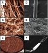
Tissue Engineering Strategies to Repair Ruptured Achilles Tendons. Tissue engineering strategies utilizing 3D scaffolds have been engineered to integrate between ruptured ends of Achilles tendons. (A–C) The collagen-PDS implant engineered demonstrated fibril-like characteristics when viewed in the transverse direction (A, scale: 20μm) and porous morphology with fibers connected by cross links in the transverse direction (B, scale: 20μm). In vivo and ex vivo experiments demonstrated that collagen-PDS implants recapitulate the structural, compositional, and mechanical properties of control tendons, and show capacity for good integration. (C) SEM showing the entire collagen-PDS implant (scale: 1.2 mm). (C–F) In a separate approach, poly(3-hydroxybutryrate-co-3-hydroxyhexanoate) (PHBHHx) was evaluated as a potential scaffold for rat Achilles tendon healing. SEM images demonstrated that the fibers (D; scale: 200× magnification)used to construct the PHBHHx tubular scaffold also contained surface pores (E; scale: 500×). (F) SEM showing entire PHBHHx implant. Once implanted, these scaffolds showed comparable mechanical properties to rat tendon, no secondary immune response, evidence of tissue remodeling and cell alignment, and a return to normal animal load bearing. Panels A–C: Image reproduced with permission from Meimandi-Parizi et al. (2013). Panels D–F: Image reproduced with permission from Webb et al. (2013).

References

    1. Kotian SR, Sachin KS, Bhat KM. Bifurcated plantaris with rare relations to the neurovascular bundle in the popliteal fossa. Anat Sci Int. 2013;88:239–241. - PubMed
    1. Fukashiro S, Komi PV, Jarvinen M, Miyashita M. In vivo Achilles tendon loading during jumping in humans. Eur J Appl Physiol Occup Physiol. 1995;71:453–458. - PubMed
    1. Maffulli N. Rupture of the Achilles tendon. J Bone Joint Surg Am. 1999;81:1019–1036. - PubMed
    1. Jarvinen TA, Kannus P, Maffulli N, Khan KM. Achilles tendon disorders: etiology and epidemiology. Foot Ankle Clin. 2005;10:255–266. - PubMed
    1. Nyyssonen T, Luthje P, Kroger H. The increasing incidence and difference in sex distribution of Achilles tendon rupture in Finland in 1987–1999. Scand J Surg. 2008;97:272–275. - PubMed

LinkOut - more resources